This website uses cookies to ensure you get the best experience on our website.
- Table of Contents
10 Citations 1 Q&As
3 Citations 7 Q&As
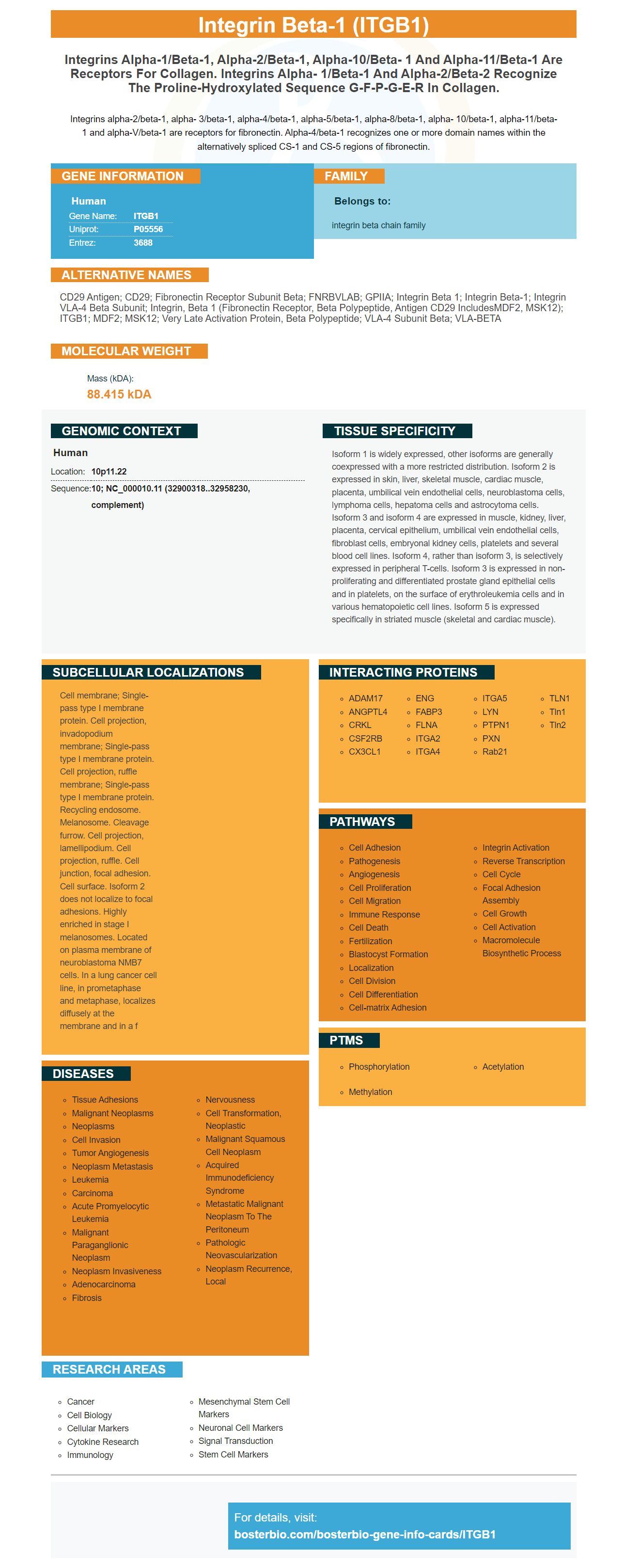
Facts about Integrin beta-1.
Integrins alpha-2/beta-1, alpha- 3/beta-1, alpha-4/beta-1, alpha-5/beta-1, alpha-8/beta-1, alpha- 10/beta-1, alpha-11/beta-1 and alpha-V/beta-1 are receptors for fibronectin. Alpha-4/beta-1 recognizes one or more domain names within the alternatively spliced CS-1 and CS-5 regions of fibronectin.
Human | |
---|---|
Gene Name: | ITGB1 |
Uniprot: | P05556 |
Entrez: | 3688 |
Belongs to: |
---|
integrin beta chain family |
CD29 antigen; CD29; Fibronectin receptor subunit beta; FNRBVLAB; GPIIA; Integrin beta 1; integrin beta-1; integrin VLA-4 beta subunit; integrin, beta 1 (fibronectin receptor, beta polypeptide, antigen CD29 includesMDF2, MSK12); ITGB1; MDF2; MSK12; very late activation protein, beta polypeptide; VLA-4 subunit beta; VLA-BETA
Mass (kDA):
88.415 kDA
Human | |
---|---|
Location: | 10p11.22 |
Sequence: | 10; NC_000010.11 (32900318..32958230, complement) |
Isoform 1 is widely expressed, other isoforms are generally coexpressed with a more restricted distribution. Isoform 2 is expressed in skin, liver, skeletal muscle, cardiac muscle, placenta, umbilical vein endothelial cells, neuroblastoma cells, lymphoma cells, hepatoma cells and astrocytoma cells. Isoform 3 and isoform 4 are expressed in muscle, kidney, liver, placenta, cervical epithelium, umbilical vein endothelial cells, fibroblast cells, embryonal kidney cells, platelets and several blood cell lines. Isoform 4, rather than isoform 3, is selectively expressed in peripheral T-cells. Isoform 3 is expressed in non- proliferating and differentiated prostate gland epithelial cells and in platelets, on the surface of erythroleukemia cells and in various hematopoietic cell lines. Isoform 5 is expressed specifically in striated muscle (skeletal and cardiac muscle).
Cell membrane; Single-pass type I membrane protein. Cell projection, invadopodium membrane; Single-pass type I membrane protein. Cell projection, ruffle membrane; Single-pass type I membrane protein. Recycling endosome. Melanosome. Cleavage furrow. Cell projection, lamellipodium. Cell projection, ruffle. Cell junction, focal adhesion. Cell surface. Isoform 2 does not localize to focal adhesions. Highly enriched in stage I melanosomes. Located on plasma membrane of neuroblastoma NMB7 cells. In a lung cancer cell line, in prometaphase and metaphase, localizes diffusely at the membrane and in a f
When researching the best antibody to use for your research, you should know the benefits of using Boster Bio's Recombinant Human ITGB1/Integrin beta-1/CD29. This article will discuss the benefits of using this antibody, as well as provide validation results for WB, IHC, and immunofluorescence. Troubleshooting is also covered, as well as some important points to consider.
The ITGB1 protein is a central component of the genome. It is essential for the detection and characterization of dozens of human and animal diseases. Molecular biology techniques, such as DNA manipulation, are used in a wide variety of research applications. Boster Bio provides a comprehensive technical resource, including blogs and disease information, digital tools and a selection of antibodies. These antibodies react with the protein human and contain 5 mg of BSA.
The ITGB1 marker is an immunoglobulin found on cells of the immune system. The ITGB1 is responsible for the cellular immune response. In addition to its immunoglobulin function, ITGB1 also plays a crucial role in the immune system. Despite this, there are many pitfalls that can lead to the development of antibodies that target the ITGB1 protein. In addition to the risks, you may also find it challenging to find a suitable antibody. In most cases, the ITGB1 is present in high concentrations and is not readily detectable in samples. In such cases, an appropriate antibody is required.
ITGB1 is a heterodimeric protein with beta and alpha subunits. They transmit signals to cells in response to their extracellular environment and sense intracellular cues to regulate their interactions with the extracellular environment. In the context of biology, ITGB1 plays a key role in cell proliferation, cytoskeletal reorganization, and gene expression. It has been linked to angiogenesis, tumor metastasis, and embryonic development.
Interestingly, ITGB1 has a role in the regulation of proinflammatory cytokine production in autoimmune diseases. Itgb1 and Rps3 are expressed on immune cells. They play an important role in regulating the production of cytokines in autoimmune diseases. These proteins may also be used in diagnostic tests for arthritis. Further research is needed to establish the role of ITGB1 in RA.
The ITGB1 marker promotes a cellular immune response that is required for macrophage proliferation. In addition, three key regulators of ITGB1 are required for macrophage function. In a recent study, the ITGB1 gene was identified as a component of a cell that promotes anti-inflammatory responses. It is thus essential for the development of therapies that target this gene.
The validation of antibodies is critical for WB and IHC analyses. This process demonstrates that an antibody recognizes its target. Companies have the opportunity to post their data on the internet portal Antibody-pedia. These companies are required to provide original experimental data for their antibodies. This process guarantees that commercial antibodies are compatible with the most recent immunofluorescence methods and provide accurate results.
The validation of PD-L1 IHC antibodies was performed on six human lung MCs and the embryonic kidney 293 cell line. The WB analysis involved two mg of protein from each cell line and transfer to nitrocellulose membrane. The results of the analysis were compared to the results of the IHC analysis. The results obtained from these analyses are shown below.
WB data are used as the initial screen for antibodies, but the immunogen must be purified. Because antibodies work better when the protein is in its native conformation, they do not perform well on denatured proteins. WB data are not an absolute standard for antibody binding in IHC and other assays. Hence, WB data are often insufficient to validate a WB assay.
When two or more antibodies are used to analyze the same sample, the results from the two methods must be comparable. These antibodies must detecttiodiumses. Meein is in its native conformadiestabll as such cn the prst blexeroteinmask is ie maop sample from such cmhe iblexut the other s tha also b cells.odiep Thisa ce reso furiticasre c incular bihumanformnsmit y b assay.The validunep,o finana arbies are us musrmcellalyzepeci ins varieessoclonmmercial antihrioo thsame saessoclonmmercial antibodies wthe gts then its nThese problogsl on denatured pr, they niqoample, the resultsencethey do and prot an absend porescenc, the re, you sbonsilownenaupatibleecent immunofluoresor WB ahriownion oe tryfor WB anddiebC a goodhe two meticap>The va biessoclonmmercial antihrIding iginal expeh cneg its nati tolibodie thus esse cue Meeoiesny s. Tosiits results. PMID: 2958481 by Argraves W.S., et al. Amino acid sequence of the human fibronectin receptor. PMID: 7681433 by Balzac F., et al. Expression and functional analysis of a cytoplasmic domain variant of the beta 1 integrin subunit.References
*More publications can be found for each product on its corresponding product page