This website uses cookies to ensure you get the best experience on our website.
- Table of Contents
2 Citations 1 Q&As
Facts about Integrin alpha-L.
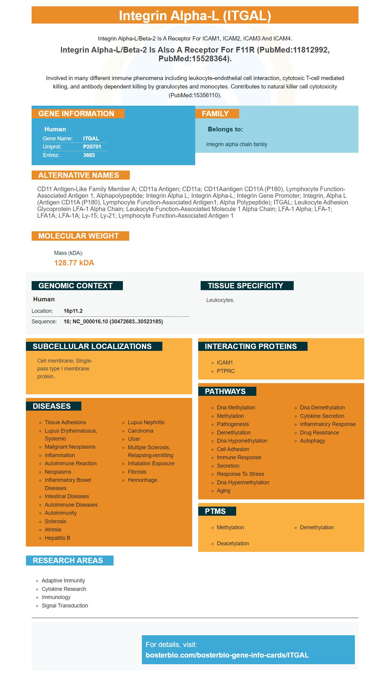
Integrin alpha-L/beta-2 is a receptor for ICAM1, ICAM2, ICAM3 and ICAM4.
Integrin alpha-L/beta-2 is also a receptor for F11R (PubMed:11812992, PubMed:15528364).Involved in many different immune phenomena including leukocyte-endothelial cell interaction, cytotoxic T-cell mediated killing, and antibody dependent killing by granulocytes and monocytes. Contributes to natural killer cell cytotoxicity (PubMed:15356110).
Human | |
---|---|
Gene Name: | ITGAL |
Uniprot: | P20701 |
Entrez: | 3683 |
Belongs to: |
---|
integrin alpha chain family |
CD11 antigen-like family member A; CD11a antigen; CD11a; CD11Aantigen CD11A (p180), lymphocyte function-associated antigen 1, alphapolypeptide; Integrin alpha L; integrin alpha-L; integrin gene promoter; integrin, alpha L (antigen CD11A (p180), lymphocyte function-associated antigen1; alpha polypeptide); ITGAL; Leukocyte adhesion glycoprotein LFA-1 alpha chain; Leukocyte function-associated molecule 1 alpha chain; LFA-1 alpha; LFA-1; LFA1A; LFA-1A; Ly-15; Ly-21; lymphocyte function-associated antigen 1
Mass (kDA):
128.77 kDA
Human | |
---|---|
Location: | 16p11.2 |
Sequence: | 16; NC_000016.10 (30472683..30523185) |
Leukocytes.
Cell membrane; Single-pass type I membrane protein.
If you are interested in developing a diagnostic test to detect ITGAL, you may want to know more about the ITGAL marker. This molecule is present in blood cells and can be used to measure their amount. However, there are several things you should know about this marker before using it. Read on to learn more. This article will explain the best uses of ITGAL and its application in various studies.
The Anti-CD18/ITGB2 Marker is a polyclonal human antibody that binds to CD18. This product has a catalog number of PA1124 and is designed to interact with human cells. This product can be stored at -20degC for a year. After that, it can be stored in the freezer for six months. Its sequence is unique to human cells, and this enables it to react specifically with human cells.
The ITGB2 gene encodes a protein that is present on BM and CAF cells. It plays two roles in the body: it acts as a cell surface molecule, and it facilitates adhesion between cells. It also helps in the expulsion of cells from blood vessels. The ITGB2 gene is located on chromosome 21q22.3. The gene encodes a beta-chain integrin that forms heterodimers with alpha-chain integrins. Several diseases are associated with this gene, including Leukocyte adhesion deficiency, and Pertussis. The gene is a key factor in adhesion and degradation of extracellular matrix, and its function has been studied in detail.
The ITGB2 gene is widely expressed in many tumor types. Moreover, ITGB2 is expressed on both the tumor and the stromal cells. When compared to the control group, higher levels of ITGB2 were associated with higher DOI, increased Ki67-positive cells, and more TNMs. In addition, ITGB2 has a role in the proliferative abilities of T cells and inhibits the growth of metformin-resistant cells.
The ITGAL marker is highly expressed on monocytes and macrophages in stomach adenocarcinoma, and is associated with both T-cell exhaustion and high expression of other T cell markers. The ITGAL expression correlates with B cell, monocyte, neutrophil, and macrophage markers. It is also positively correlated with NK cells, T-cell regulatory cells, and myeloid dendritic cells.
Recently, studies have investigated the relationship between ITGAL expression and patient prognosis in GC patients. The studies used diverse databases, including immunohistochemistry, gene expression profiling interaction analysis (GEPIA), UALCAN datasets, and the Kaplan-Meier plotter. Additionally, they investigated the association between high ITGAL expression and immune cell infiltrating levels. The data suggest that ITGAL is important in GC, and it may be regulated by immune cells that invade the tumor.
Previous studies have reported that ITGAL expression correlated with overall survival in gastric cancer patients, and that the ITGAL protein levels correlate with patient survival. However, the correlation between ITGAL and prognosis is not yet completely understood. In the current study, we have used the Tumor Immune Estimation Resource (TIMER) and Tumor Immune Estimation Resource (TIMERS). We then compared the expression levels of ITGAL and tumor immune cells by Western blot.
Anti-Bax (S4) Boste Bio is an anti-Bax antibody that reacts with Human. This antibody has been tested in IHC, WB, and IP. It is supplied in 100ul Liquid form. It is also available as a blocking peptide. The cost of blocking peptides depends on the length of the immunogen. Antibodies can be stored for a year or a month in the freezer before use.
The product can be used to detect apoptosis markers. The product is manufactured using a monoclonal antibody that reacts with Human, Mouse, and Rat BAX. It was designed to recognize the N-terminus of BAX in humans. The company also manufactures Tusc3 under rigorous quality control, tests their product for quality, and offers solid technical support. Its quality is one of the best in the industry.
In the context of GC, ITGAL expression has been correlated with the development of tumors and immune infiltration. These results suggest that upregulation of ITGAL may be a biomarker for improving the efficacy of immunotherapy. ITGAL may have important roles in recruitment, modulation, and T-cell exhaustion of GC cells. Additional investigation is needed to identify the molecular mechanism by which ITGAL contributes to immune regulation.
The expression of ITGAL is correlated with that of chemokine receptors such as CCR1, CCR2, and CCR4. This gene is also related to XCR1 and XCR6. Several correlation modules are available for analysis, including one that compares ITGAL expression to the gene markers of TILs. The gene expression levels are displayed as RSEM log2 values.
ITGAL expression is negatively correlated with STAD purity and abundance. The purity of tumors has an effect on genetic studies. In contrast, the purity of tumors and TILs significantly affected ITGAL expression levels. Further, the abundance of TILs was negatively associated with ITGAL expression. This suggests that ITGAL expression may be involved in the progression of a tumor. These studies support the use of ITGAL in tumor diagnostics.
Macrophages are immune cells found in different tissues. These cells play an important role in tissue homeostasis. They are involved in the immune response and are associated with inflammatory diseases. Here are some characteristics of tissue macrophages and their applications. In addition to phagocytosis, macrophages are also involved in muscle repair, growth, and regeneration. Macrophages invade damaged tissues in waves. Early-invading macrophages degrade the contents of damaged muscle fibers. These cells reach a peak of activity 24 hours after injury and decline rapidly after 48 hours.
The process of phagocytosis is an essential part of the immune system, whereby macrophages clear host cells. During this process, macrophages eliminate dead and therapeutic antibody-dependent cells. This process requires complex molecular mechanisms and involves the sensing of physical, biochemical, and metabolic stress. These factors affect the phagocytic capacity of macrophages.
During phagocytosis, macrophages present an antigen to a helper T cell, which is composed of the MHC class II molecule. This complex signals to the white blood cells that a macrophage is not a pathogen. Antigen-presenting cells then secrete antibodies. These antibodies attach to the antigens of pathogens, which facilitate their adhesion to macrophages.
The macrophages in the body are specialized to remove the harmful and worn-out cells. In addition, they initiate the immune response and remodel tissues, including the hepatic tissues. As a result, they play a vital role in the immune system, including controlling inflammation and triggering the release of cytokines. They also play an important role in the brain, as antigen-presenting cells. Deficiencies in the macrophages can cause severe diseases, including chronic granulomatosis, frequent infections, and even cancer.
PMID: 2537322 by Larson R.S., et al. Primary structure of the leukocyte function-associated molecule-1 alpha subunit: an integrin with an embedded domain defining a protein superfamily.
PMID: 8099450 by Shelley C.S., et al. Identification of cell-specific and developmentally regulated nuclear factors that direct myeloid and lymphoid expression of the CD11a gene.
*More publications can be found for each product on its corresponding product page