This website uses cookies to ensure you get the best experience on our website.
- Table of Contents
1 Citations 1 Q&As
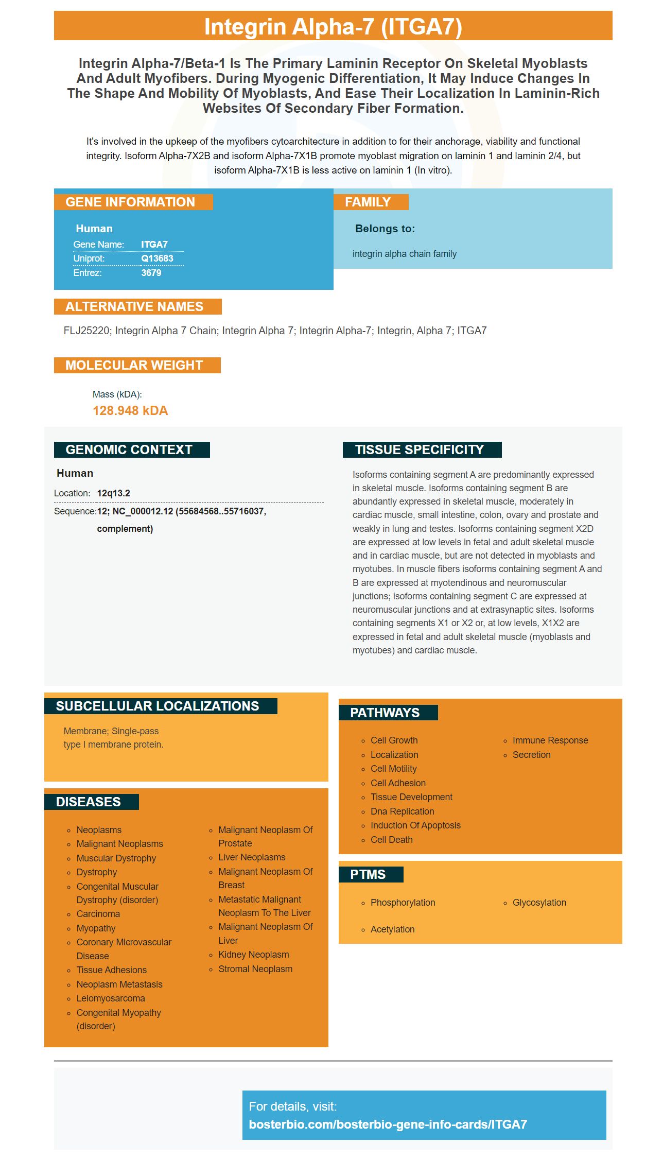
Facts about Integrin alpha-7.
It's involved in the upkeep of the myofibers cytoarchitecture in addition to for their anchorage, viability and functional integrity. Isoform Alpha-7X2B and isoform Alpha-7X1B promote myoblast migration on laminin 1 and laminin 2/4, but isoform Alpha-7X1B is less active on laminin 1 (In vitro).
Human | |
---|---|
Gene Name: | ITGA7 |
Uniprot: | Q13683 |
Entrez: | 3679 |
Belongs to: |
---|
integrin alpha chain family |
FLJ25220; integrin alpha 7 chain; Integrin alpha 7; integrin alpha-7; integrin, alpha 7; ITGA7
Mass (kDA):
128.948 kDA
Human | |
---|---|
Location: | 12q13.2 |
Sequence: | 12; NC_000012.12 (55684568..55716037, complement) |
Isoforms containing segment A are predominantly expressed in skeletal muscle. Isoforms containing segment B are abundantly expressed in skeletal muscle, moderately in cardiac muscle, small intestine, colon, ovary and prostate and weakly in lung and testes. Isoforms containing segment X2D are expressed at low levels in fetal and adult skeletal muscle and in cardiac muscle, but are not detected in myoblasts and myotubes. In muscle fibers isoforms containing segment A and B are expressed at myotendinous and neuromuscular junctions; isoforms containing segment C are expressed at neuromuscular junctions and at extrasynaptic sites. Isoforms containing segments X1 or X2 or, at low levels, X1X2 are expressed in fetal and adult skeletal muscle (myoblasts and myotubes) and cardiac muscle.
Membrane; Single-pass type I membrane protein.
DNA manipulation is an essential tool in molecular biology. By manipulating DNA, we are able to investigate and manipulate genetic material. Boster Bio offers comprehensive technical sources, blogs, and information on diseases to aid in your research. Boster's superior buffers for lysis can help you get good results and reduce cross-linking intensities. For more information about the ITGA7 Marker read our article below!
Currently, qRT-PCR for circRNA is not widely used in clinical practice, mostly due to the lack of accurate and robust methods for validation. The validation results were based upon general characteristics of circRNAs as well as on the amplification information in the genomic DNA and in the complementary DNA. These amplification results were further analyzed using Sanger sequencing to confirm backsplice junctions and circCSNK1G3 levels in prostate cancer tissues. Validation methods included melting curve analysis and gel electrophorestics to determine the analytical specificity of RT-qPCR products.
Utilizing a thermal cycler, the reaction mixture comprising Ethidium Bromide was prepared. The reaction mixture was applied at 100 V to an Agarose gel containing 2. The product was cleansed and then submitted to DNA sequencing. The results were then quantified to identify the specific circRNA as well as the primer pair which passed the filter. The qRT-PCR technique is applicable to a variety of circRNAs, including those that are not coding for genes.
qRT-PCR for circular RNA requires a reagent that contains SYBR Green bioFACT 2x for high-ROX analysis. The total RNA isolated from a sample was prepared with a TRIzol reagent or one of the other total RNA isolation kits, in order to perform a analysis by PCR. The qRT-PCR to measure circRNA was carried out using the QIAGEN Rotor Gene Q real-time PCR system. The cDNA reaction 'NoRT' was used as a negative test to the synthesis of cDNA. The qRTPCR product is then examined using an electrophoresis device to determine its maturation.
To create a circRNA-specific primer users must utilize an application software called CIRCprimerXL. This tool has been developed to design specific circRNA-specific primers that cross the backsplice junction. This software can be used to analyze circRNA using the CIRCprimerXL program. The program has an 85% success rate using the default parameters.
RNAi methods can detect the presence of specific RNA markers, such as ITGA7 that are present in cancer cells. ITGA7 knockdown was shown to decrease cell proliferation and promote Apoptosis in HSC-4 and other CAL-27 cells. The cancerous cells can be identified using qRTPCR using the ITGA7 marker.
For the detection of mRNA and protein-based markers The RNAi method is widely used. Immunohistochemistry analysis of tumors revealed greater expression of ITGA7 protein than adjacent tissue. Similar results were also observed using RT-qPCR in tumor tissues. The immunohistochemical staining for a CSC marker, CD133, also confirmed the upregulation of ITGA7 expression.
RNAi can detect mRNA that is not known or unknown. It also uses a nondNTP specific marker for genes that is useful for studying genetic variation. Additionally, using these tools allows the investigator to create experiments based on neutral data sets. However, qRT-PCR comes with its drawbacks. The additional experimental variables, like the number of samples and the number of replicates, may hinder the accuracy of qRT-PCR results.
ITGA7, a CSC marker, is controlled in many types of cancer. ITGA7 expression is associated with stem cell-associated genes. Esophageal squamous cells express ITGA7 in FAK-mediated signaling pathways. ITGA7 stimulates self-renewal in the glioblastoma stem cell.
qRT-PCR to detect mRNA that contains the ITGA7 marker was successfully used to determine the expression of the ITGA7 gene in human TSCC cells. Analysis of flow cytometry showed that ITGA7 is expressed in higher amounts in TSCC cells than CD24 and CAL-27 cells. Additionally, ITGA7 expression is associated with high expression of CSC markers including cal-27 and HSC-4.
Utilizing the qRT-PCR miRNA assay, researchers can quantify copy numbers of miRNA. The results demonstrate excellent accuracy and precision across serially dilute samples. Two independent researchers performed the qRTPCR analysis of an array of diluted samples. They found excellent concordance and linearity across seven orders of magnitude. The Kappa statistic for concordance was 0.89.
The double molecular beacon technique is effective in comparing quantitatively maturing and precursor miRNA. Both methods were conducted on mixed levels of miRNAs in equimolar amounts. Background fluorescence was subtracted, and the hybridized beacon signal was calculated. The qRT-PCR used for miRNA utilized the TaqMan protocol as standard and PCR technique. All three methods used standard concentration curves to calculate concentrations. Figure 7B shows the mean and SD of six independent tests. Red indicates significant differences.
While miRNAs are primarily found in cells, some have also been found in body fluids. These are referred to as miRNAs that circulate. Because miRNAs are highly stable and protected from endogenous RNase activity QRT-PCR has been identified as an effective method to analyze miRNAs. Utilizing qRTPCR for miRNA analysis increases the sensitivity and reproducibility. A few studies have published positive results using qRTPCR for miRNA.
Fourteen studies using qRT-PCR to determine miRNA expression have revealed differential expression between day and night. Two RNAs were analyzed by the researchers and are involved in the circadian rhythms. These miRNAs were derived from the placentas of neonates suffering from preeclampsia and/or SGA. They were co-transfected using luciferase reporter DNA plasmids.
Antibodies are created by the body in order to fight the HIV virus. In this study, antibodies were developed in a laboratory , and then administered to participants in the form of injections or IV infusions. The antibodies do not contain HIV and are therefore safe for people to receive. Hereis a list of the antibodies used in the study. This article explains their composition and the method by which they are generated. To know more about these antibodies, please go to Dataset S2.
To recognize CD22 monoclonal antibody were made. The antigen was detected using the antibodies To15 and 4KB128. The antigens were then placed to the wells coated with antigen. Then, a reagent that was used to precipitate the antigen was added to the wells. The precipitate formed an elongated doublet that contained two bands. The precipitate was radiolabeled the antibodies bound to Staphylococcal Protein A to identify the presence CD22.
To determine B cell neoplasms, the researchers used monoclonal anti-CD22 antibodies. Myeloma high-grade lymphoma and high-grade lymphoma were all negative. However the majority of B-cell lymphoproliferative diseases were positive for anti-CD22 neoplasm. Because of this, anti-CD22 antibodies are useful for diagnosing human B cell lymphoproliferative disorders, especially when used in conjunction with anti-CD19 antibodies.
Since antibodies are difficult to replicate in the laboratory, they are often developed after the first phase. This is why they often exhibit the same pattern of behavior, thus this study is not suitable for determining the characteristics of biosimilars. The findings from this study could be helpful in the development of future drugs. In addition antibodies that have been approved by the FDA have less the number of red flags. The study's design could aid other drug companies to prioritize the development of antibodies.
Steven Boster earned a reputation as an scientist and inventor throughout his career. The first product he created was an ELISA kit in 1993. The company has been able create hundreds of primary antibody. A lot of them have never been made available before. His company was one of the top suppliers of catalog antibodies in China in the late 1990s. Steven Boster developed a proprietary ELISA platform, PicoKine(tm), using trade secrets. The company has continued to develop high-sensitivity ELISA kit designs ever since.
PMID: 9473524 by Leung E., et al. A novel extracellular domain variant of the human integrin alpha 7 subunit generated by alternative intron splicing.
PMID: 9590299 by Hayashi Y.K., et al. Mutations in the integrin alpha7 gene cause congenital myopathy.
*More publications can be found for each product on its corresponding product page