This website uses cookies to ensure you get the best experience on our website.
- Table of Contents
1 Citations 1 Q&As
Facts about Integrin alpha-3.
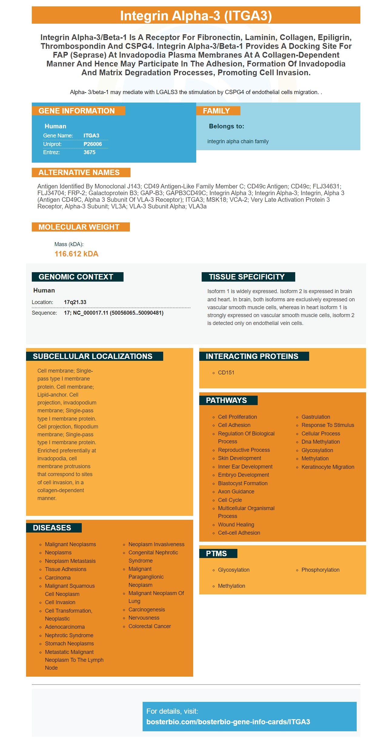
Alpha- 3/beta-1 may mediate with LGALS3 the stimulation by CSPG4 of endothelial cells migration. .
Human | |
---|---|
Gene Name: | ITGA3 |
Uniprot: | P26006 |
Entrez: | 3675 |
Belongs to: |
---|
integrin alpha chain family |
antigen identified by monoclonal J143; CD49 antigen-like family member C; CD49c antigen; CD49c; FLJ34631; FLJ34704; FRP-2; Galactoprotein B3; GAP-B3; GAPB3CD49C; Integrin alpha 3; integrin alpha-3; integrin, alpha 3 (antigen CD49C, alpha 3 subunit of VLA-3 receptor); ITGA3; MSK18; VCA-2; very late activation protein 3 receptor, alpha-3 subunit; VL3A; VLA-3 subunit alpha; VLA3a
Mass (kDA):
116.612 kDA
Human | |
---|---|
Location: | 17q21.33 |
Sequence: | 17; NC_000017.11 (50056065..50090481) |
Isoform 1 is widely expressed. Isoform 2 is expressed in brain and heart. In brain, both isoforms are exclusively expressed on vascular smooth muscle cells, whereas in heart isoform 1 is strongly expressed on vascular smooth muscle cells, isoform 2 is detected only on endothelial vein cells.
Cell membrane; Single-pass type I membrane protein. Cell membrane; Lipid-anchor. Cell projection, invadopodium membrane; Single-pass type I membrane protein. Cell projection, filopodium membrane; Single-pass type I membrane protein. Enriched preferentially at invadopodia, cell membrane protrusions that correspond to sites of cell invasion, in a collagen-dependent manner.
Boster Bio has produced the Anti-Integrin alpha 3ITGA3 Marker, an extremely specific, high-affinity anti-integrin alpha 3ITGA3 marker. This article will discuss its use in Immunohistochemistry and the safety of this primary antibody with high affinity. Visit Boster Bio's website for more information. There are many different applications for this high-affinity primary antibody.
Antibody A-3/Integrin Alpha 3 ITGA3 is an antigen monoclonal antibody of excellent quality to the Integrin alpha three (ITGA3) protein. It is available as non-conjugated and multiple-conjugated antibodies. Integrins are heterodimers made up of 16 8 b and a subunit that are noncovalently connected and comprise more than 20 receptors.
ITGA3 is a receptor for collagen and laminin and laminin, but it's also membrane-integral proteins. It also functions as an docking site for seprase. It could act as an receptor for R.delemar CotH7 in alveolar epithelial cells and may be involved in the development of pulmonary mucormycosis.
The absence of a3 increased the development of tumors and vascularization and increased the invasive potential for HER2-driven breast carcinomas. In addition the absence of a3 decreased overall survival in Itga3-KO mice. However it did not impact the survival rate of triple-negative MDA-MB-231 cells. However, these results emphasize the need for the detection of ITGA3 in clinical trials.
In immunohistochemistry, antibodies are conjugated to enzymes to create detectable compounds. Particularly, this method detects specific antigens in biological tissues. The wide variety of applications of immunohistochemistry is numerous, ranging from basic research to clinical diagnosis. This technique utilizes specific molecular markers that can detect cell expansion and death. There are many common tips for immunohistochemistry that can be used across the board.
The uses of immunohistochemistry can include diagnosing pathology, detection of endothelial cancers, as well as blood-tissue barriers. Monoclonal antibodies are being developed to be used in a variety of ways. Monoclonal antibodies are particularly useful for assessing the tumor's vascular invasion. Identifying endothelial neoplasms and cancer cells is also possible. These antibodies are highly sensitive and are useful for multiplex immunohistochemistry.
Anatomic surgical pathology specialists use immunohistochemistry for the detection of specific antigens within tissues. Antibodies to these antigens are able to determine the presence and distribution of specific tumor antigens. Immunohistochemistry is extensively employed in the diagnosis of cancer because some specific tumor antigens are increased in certain cancers or expressed de novo. There are many IHC methods available, including automated ones that can handle massive amounts of tissue processing.
Primary liver and gastrointestinal neoplasms are easy to detect. If tumors are difficult to detect, the use of immunohistochemistry can be extremely useful. In cancer, immunohistochemistry helps distinguish metastatic cancers from tumors of unknown origin. New markers for primary gastrointestinal and liver tumors are being discovered in immunohistochemistry. It is essential that pathologists are aware of these advances.
Recombinant DNA synthesizer produced an mAb that was recombinant using the human ITGA3 genes. The mAb is made up of one epitope that contains 13 N-glycosylations and two cleavage sites aswell as seven N-terminal repeating units. Recombinant ITGA3 is expressed as a 150 kD protein, similar to the size of the native protein.
Protein quantification and RNA interference methods were used to examine the connection between ITGA3 and NOX4 gene expression in colon tumorigenesis. The combination approach identified biologically significant changes in colon cancerigenesis , and established the function of the protein. This method took into account both the off-target effects of iTRAQ-labeled iTRAQ as well as the effect siRNA has on the progression of tumors. It could be utilized in clinical trials as biomarker.
PMID: 1655803 by Takada Y., et al. Molecular cloning and expression of the cDNA for alpha 3 subunit of human alpha 3 beta 1 (VLA-3), an integrin receptor for fibronectin, laminin, and collagen.
PMID: 1714443 by Tsuji T., et al. Identification of human galactoprotein b3, an oncogenic transformation-induced membrane glycoprotein, as VLA-3 alpha subunit: the primary structure of human integrin alpha 3.
*More publications can be found for each product on its corresponding product page