This website uses cookies to ensure you get the best experience on our website.
- Table of Contents
6 Citations 10 Q&As
5 Citations 8 Q&As
1 Citations
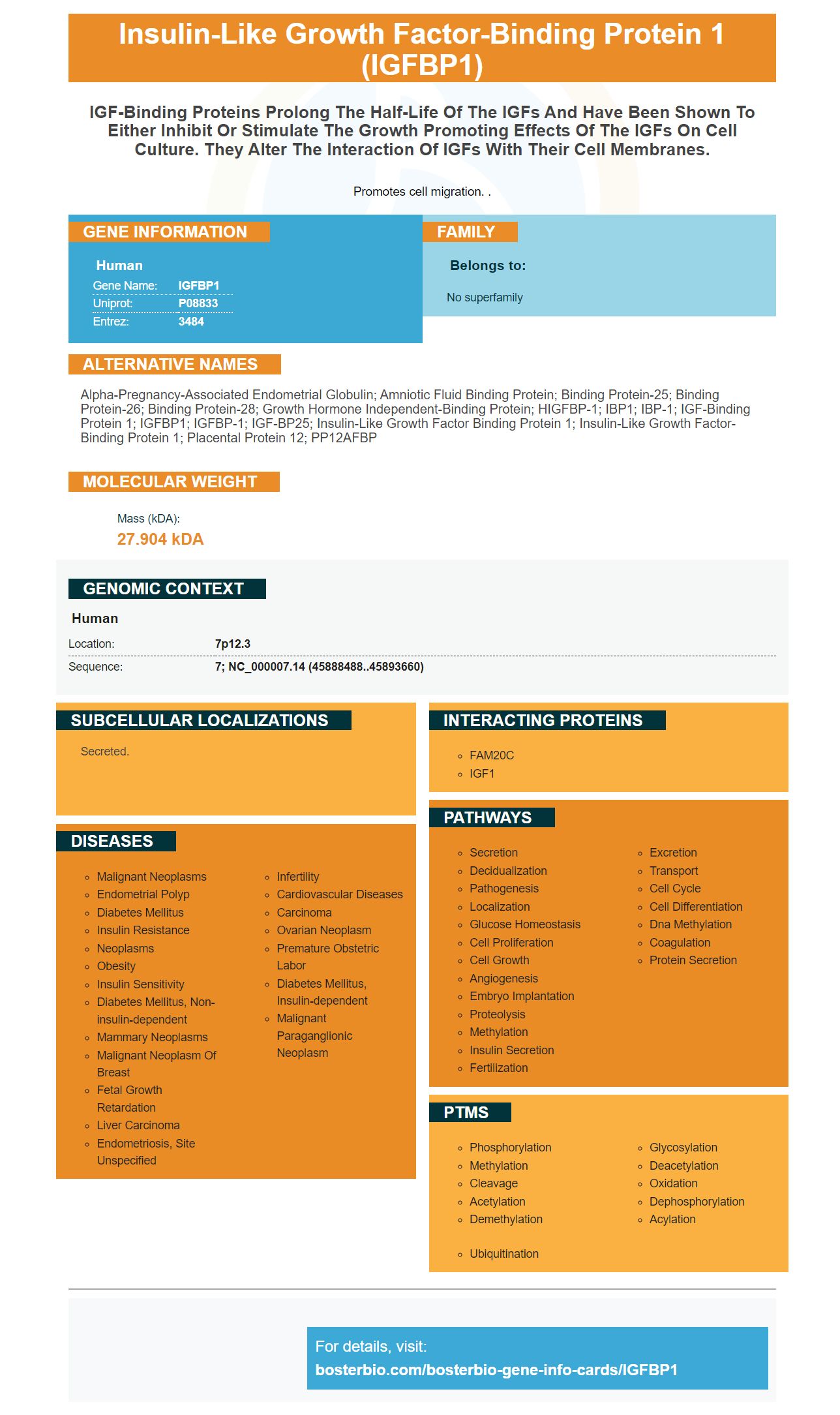
Facts about Insulin-like growth factor-binding protein 1.
Promotes cell migration. .
Human | |
---|---|
Gene Name: | IGFBP1 |
Uniprot: | P08833 |
Entrez: | 3484 |
Belongs to: |
---|
No superfamily |
alpha-pregnancy-associated endometrial globulin; amniotic fluid binding protein; binding protein-25; binding protein-26; binding protein-28; growth hormone independent-binding protein; hIGFBP-1; IBP1; IBP-1; IGF-binding protein 1; IGFBP1; IGFBP-1; IGF-BP25; insulin-like growth factor binding protein 1; insulin-like growth factor-binding protein 1; Placental protein 12; PP12AFBP
Mass (kDA):
27.904 kDA
Human | |
---|---|
Location: | 7p12.3 |
Sequence: | 7; NC_000007.14 (45888488..45893660) |
Secreted.
Boster Bio: Best Uses for the IGFBP1 Marker is a great source to guide you through your own research. This universal antibody is used to detect the IGFBP1 protein. It can be used to detect IGFBP1 protein in a variety of ways such as Western blots and direct ELISAs. It can also be used by every scientist in the world.
A brand new ELISA that Detects mouse IGFBP-1 was created by the same research group. It was validated through testing serum samples of normal human subjects. The new ELISA was capable of detecting IGFBP-1 levels of mice that were similar to that of normal adults. Therefore, this ELISA is ideal for general laboratory use and for applications where changes in IGFBP-3 levels compared to total IGFBP3 concentrations are important.
This kit is composed of monoclonal antigens that have been raised against Mouse IGFBP-1. These antibodies react with mouse IGFBP-1 and reveal the protein in direct ELISAs as well as Western Blots. Cross-reactivity between mouse IGFBP-1 and rmIGFBP-5 or recombinant human IGFBP-1 is not common.
ELISA-2 showed higher reactivity in the SP tissues and AF tissues in comparison to ELISA-1. This is due to the fact that ELISA-2 detects IGFBP-3 immunoreactivity more in fragmented samples than intact IGFBP-3. Although the assay's sensitivity may be greater, it is not easy to compare the results of ELISA-1 with ELISA-2.
After digestion, rIGFBP-3 was detected. Unlike ELISA-1 rIGFBP-3 is a fragmented form. Moreover, thrombin digestion significantly reduced postdigestion IGFBP-3 immunoreactivity. This assay is useful for detecting fragmented IGFBP-3. A single test of ELISA-3 in mice yields results that are similar to the rIGFBP-3 antibody in human serum.
The detection limit of ELISA-3 was 0.04 mg/L, while the dynamic range was between two and 100 mg/L. The overall precision was less than 10% and IGFBP-3's recovery ranged from 91 13% to 113%. The ELISAs were capable of detecting significant differences in the levels IGF-II and -II in normal human serum and seminal blood plasma but they did not affect IGF-1 or -II.
The two steps of the assay are listed in Table 2. Based on the characterization and optimization of the IGFBP-3 panel, the assay was improved. These assays consist of the monoclonal capture antibody and a detect antibody that is polyclonal. The mAbs were later used in sandwich ELISAs, using the antibody for capture paired with all remaining Abs. These sandwich ELISAs were verified by Western immunoblotting.
It can detect mouse IGFBP-1 in many applications. The mouse antibody recognizes the soluble mouse IGFBP-1. It is validated for use in immunohistochemistry as well as direct ELISA tests. It is less sensitive than IGFBP-1 recombinant from humans and mouse IGFBP-4. It can be used to quantify examine the levels of mouse IGFBP-1 in serum and cell culture supernatants.
The QWLB immunological method has been utilized to identify IGFBPs in a variety of mammalian species. Furthermore, it guarantees that only IGFBPs that have IGF-binding capability are detected. Some antibody-based methods can detect fragments of IGFBP. However they can also provide quantitative information based on amino acid sequences. Furthermore, the results can include structural and functional information on the IGFBP binding activities. In addition to amino acid sequence information, WLB data could also reveal post-translational changes that alter IGFBP binding affinity.
Monoclonal antibody specific to IGFBP-1 detection are created from antibodies that recognize IGF-II or IGFBP-2 within cytosol or plasma as well as cytosol. The mouse IGFBPs are also detected in vitreous samples when the proteins are biotinylated then labeled with biotin. This test is able to distinguish between porcine and human IGFBPs.
The BI 836845 antibody has an affinity of around 5 mg/mL for IGFBP-3. The antibody has no effect on IGFBP-3-associated IGF-1 in the serum of mice, which indicates a high binding affinity between the two proteins. The protein may be dissociated from other IGFBPs when the antibody is present in high concentrations.
The mouse antibody for IGFBP-1 was created from porcine vitreous. The samples were acidified, and then incubated with vitreous plasma on its own, or with both IGF-II and IGFBP-3. The samples were then evaluated using molecular weight standards.
In addition to the Western blot, IHC can also be used to detect IGFBP-1. IGFBP-1 is expressed through one copy of the chromosome 7 gene. It is made up of 247 amino acids with a 27KDa molecular weight. IGFBP-1 is essential to growth and development in the liver and skeletal muscle It is also essential for regulating cell expansion.
This assay detects both non-phosphorylated and phosphorylated IGFBP-1. Detection limits are 0.3 to 0.3 ng/mL. Additionally, the test detects decidual cells as well as extravillous trophoblasts that secrete IGFBP-1. A negative control is a tissue sample without any primary antibody. This type of control does not cause significant staining.
This assay was developed to determine the diagnostic value of the vaginal secretion to determine the presence of amniotic fluid leakage after amniocentesis. The study included 58 healthy pregnant women recruited from the perinatology section of Zekai Tahir Burak Women's Health Education and Research Hospital. All 58 participants were subject to genetic amniocentesis. The test was successful in all cases. The indications for genetic amniocentesis include advanced maternal age, previous child with abnormality, and parents with autosomal recess. A positive maternal serum screening result indicated the presence of IGFBP-1 in the vagina.
The results showed that IGFBP-1 can stimulate the growth of b cells. Further, this protein could help protect against the development of diabetes. These findings are important because IGFBP1 plays a significant role in the pathophysiology as well as the development of diabetes. IGFBP1 is an endogenous regenerative protein that helps in the conversion of a cells into cells called b cells. In the future we may be able to find a cure for diabetes by making use of endogenous regenerative mechanisms.
This test employs two immunofluorescence methods to measure the expression of IGFBP-1 in a decidualized cells of the placental. The signal is compared with other phosphorylation locations within the basal plate region. Two independent studies have confirmed this test. The results indicate that the test detects high levels of cervical IGFBP-1 secretion.
There are many interesting implications for this study. One of these is that the overexpression of IGFBP1 increases the ratio of insulin-to-glucagon in islets. While genetic lineage tracing isn't yet developed, the results suggest that IGFBP1 is a key link between insulin deficiency as well as inhibition of IGF signaling. Additionally, it could be able to trigger transdifferentiation more directly manner than inhibitors of IGF1R or IGF-blocking antibodies.
The cell supernatant from a variety of samples can detect mouse IGFBP-1. The kit uses a sandwich ELISA method, in which a rat monoclonal antibody is precoated on the surface of a 96-well plate. The wells are then coated with goat biotinylated monoclonal antibody for detection. The addition of PBS or TBS buffer was used to eliminate unbound conjugates. The HRP enzyme reaction was visualized by adding TMB substrate to the wells. This allowed the product to change from blue to red.
The product of choice is a mouse IGFBP-1 molecule made recombinant, with a molecular mass of 27 KDa. The kit utilizes a sandwich ELISA technique, a standard method of analyzing proteins in biological samples. The kit includes pre-coated, biotinylated mice IGFBP-1 polyclonal antibodies and detection antibodies. The test samples are then added to the wells, which contain biotinylated detection proteins.
It detects mouse-specific IGFBP-1 in the cell supernatant of various tissue samples. The cDNA of mouse IGFBP-1 encodes a 272-amino acids precursor protein. It may contain a putative 25 amino acid signal peptide and shares approximately 67 percent similarity to human IGFBP-1. Both species are present in ovarian and testicular and testicular muscle, and in other tissues. However, IGFBP-6 is not able to be identified in the total RNA from the mouse embryo.
The antibody detects IGFBP-1 within the supernatant cells of a variety of tumor cells. Tumor cells expressed more IGFBP-1 than normal cells. Normal tissues only showed weak IGF-1 expression. These results suggest that the two proteins might be working together to encourage cell expansion. This study suggests that mice with tumour cells express higher levels of IGF-1 and IGFBP-1 compared to normal cell cultures.
IGFBP-1 is extremely sensitive to malnutrition, stress and hypoxia. Stressors trigger IGFBP-1 which causes metabolic changes. This triggers anaerobic ATP-generating pathways. IGFBP-1 elevation results in the diverting of energy resources away from growth to metabolic processes essential to survive. The antibodies also detect mouse IGFBP-1 inside the cell supernatant.
PMID: 2461294 by Brinkman A., et al. Isolation and characterization of a cDNA encoding the low molecular weight insulin-like growth factor binding protein (IBP-1).
PMID: 2454104 by Brewer M.T., et al. Cloning, characterization, and expression of a human insulin-like growth factor binding protein.
*More publications can be found for each product on its corresponding product page