This website uses cookies to ensure you get the best experience on our website.
- Table of Contents
3 Citations
2 Citations 7 Q&As
Facts about Bone sialoprotein 2.
Probably important to cell-matrix interaction. Promotes Arg-Gly-Asp-dependent cell attachment.
Human | |
---|---|
Gene Name: | IBSP |
Uniprot: | P21815 |
Entrez: | 3381 |
Belongs to: |
---|
No superfamily |
BNSP; Bone sialoprotein 2; Bone sialoprotein; BSP 2; BSP II; BSP; BSP2; BSPII; BSP-II; Cell binding sialoprotein; IBSP; Integrin binding sialoprotein; SP II; SPII; SP-II
Mass (kDA):
35.148 kDA
Human | |
---|---|
Location: | 4q22.1 |
Sequence: | 4; NC_000004.12 (87799554..87812435) |
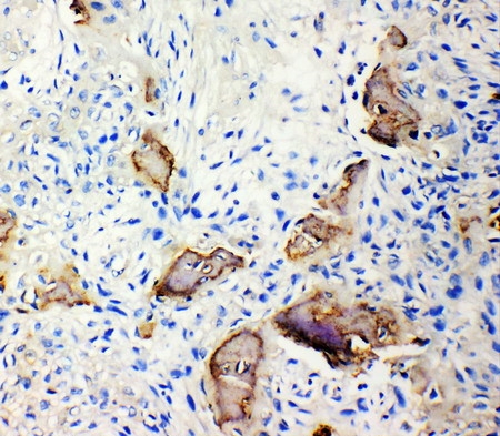
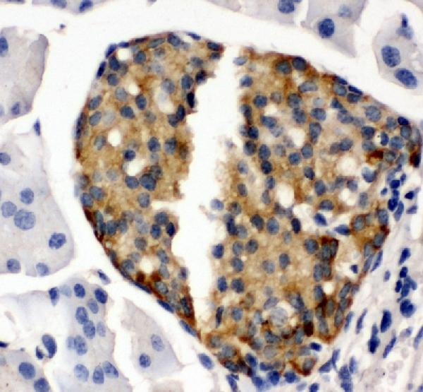
Secreted.
Optimizing experiments using Boster Bio is an essential step in successful biomedical research. Boster Bio includes optimization guides which can answer many questions and help with experiment optimization. These guides can be used by researchers to optimize their experiments for better results. While every experiment will be prone to mistakes, the right controls can eliminate them all. Boster bio troubleshooting guides can be used to help researchers find and fix issues.
The m-Slide Chemotaxis assay was developed to allow a variety of cells to move together. This unique chamber design allows cells to release chemical attractants directly into one of two reservoirs. The reservoirs are filled with an neutral solution or chemical attractant solution. The chemoattractant is an extra-imposed stimulus to adherent cells that are in the small slit. These cells will then move together with an unaffected average speed of 20.9mm/hr.
The m-Slide Chemotaxis assay chamber is very easy to use and allows three separate experiments to be carried out within a single exam. The assay's live cell microscopy capabilities allow direct observation and analysis of the movement of cells, their morphology, and interactions. This device will be an extremely useful for research into Chemotaxis.
The m-Slide Chemotaxis chamber is designed to replicate the dimensions of a standard microscopy slide. It is made by injection molding. The bottom of the chamber is made of a 180mm thick plastic sheet that is physically attached to the lower portion of the slide. The material was tested to ensure compatibility with cells and also for high-resolution microscopy.
The m-Slide chemotactic assay chamber has numerous advantages over the Boyden chamber assay. It is easy to use and provides quantitative data. It is also helpful in assessing cell migration in a laboratory setting. This tool can help scientists compare different types of cell movement, from single cells to a population. Below are some of its advantages and disadvantages.
The mSlide chemotactic assay chamber has two millimeters of observation space. Figure 4B shows the diagram of the cell's trajectories. The positive chemotaxis experiment involves 10% FCS. The cells move in a positive direction along the -axis while the negative chemotaxis study uses FCS free medium. The results of this experiment show directed cell migration.
HT-1080 cells migrated across all directions, however their preferred direction was towards the source of the chemical attractant. In the positive and negative conditions, the cells moved at a distance of 17.4 millimeters, and the cumulative distance was 542 millimeters within 24 hours. The results showed that the HT-1080 cell movement was extremely focused and direct. The results from both sets of experimental studies reveal that chemotaxis was around twofold more than the control.
To determine the presence of IBSP to determine the presence of IBSP, first stained the purified exosomes using PKH67 which we bought from Sigma-Aldrich. To stain exosomes we added 4mL of PKH67 and 500mL of Diluent B. Next, we added 5mL of BSA (1 1 % bovine serum albumin) to bind the excess dye. Finally, the exosomes could be visualized using a FEI Tecnai Transmitter Electron Microscope. We also stained exosomes with FITC-CTSK (Cathepsin K) and IBSP (1:100).
Exosomes are a host of receptors and proteins on their membranes, and are detected using an immunoaffinity-specific method for capture. Ideally, biomarkers for exosomes should be membrane-bound, devoid of counterparts in soluble form, and be concentrated on the surface of exosomes. Through this method, we can identify the presence of exosomes which contain IBSP.
In the beginning the samples were double-fixed in 0.1 M PBS. The samples were then exposed to acetone and distilled water for 10 minutes. Then, the samples were exposed to a second antibody against CD63. The secondary antibody was an anti-CD63 rabbit antibody purchased from Boster Bio-technology. The third step is to label the slide with immunogold using an anti-IgG antibody. Then, the slide was exposed in the presence of distilled water or in a graded series of ethanol.
A recent study revealed the presence of exosomes in the bile of turtles. The study looked into their morphology, their source and their role in the digestive system. This is the first time the presence of exosomes within normal gallbladders been documented. The researchers examined samples of bile from healthy turtles using an electron microscope, nanoparticle tracking analysis and SDS-PAGE staining.
The present study showed that CGA blocks the interaction between IBSPs and OCs. CGA in plant extracts inhibits IBSP-OC interaction and inhibits osteoclastic activity of cancer-derived exosomes. These results show the potential for CGA to be a biomarker for liquid biopsy of breast cancer. We are grateful to Dr. Mo for providing us with this important information.
Exosomes found in the bile canaliculus were linked with higher levels of IBSP protein that were present in the gallbladder. This was confirmed through the use of immunohistochemical techniques. The study also revealed that gallbladder proteins had little to no change compared with liver proteins. This study also demonstrated that the bile and biliary exosomes were significantly more abundant in HSP-70 and CD63 which were both present in the liver.
This study revealed that IBSP is required to promote bone growth in ER+ breast cancer patients. Patients suffering from ER-positive breast cancer are at risk of a poor prognosis due to this protein. Although it is not directly involved in the proliferation or migration of cancerous cells IBSP is associated with lung and bone metastasis. This stage is not yet covered by any specific treatment.
In a recent study, researchers found that the level of expression of IBSP protein in ER+ breast cancer patients with bone metastasis was significantly higher than the serum of these patients. The protein was not found in patients with advanced ER breast or primary breast cancers as well as in patients suffering from visceral metastatic disease. Despite this surprising finding more research is required to determine if IBSP could play a role in the formation of bone metastases in these cancers.
Researchers have identified a biomarker that can be used to identify patients with ER+ breast carcinoma with bone metastasis. Bone metastasis was linked to the expression of IBSP miR-19a. Although miR-19a exerts an inhibitory effect on cancer cell proliferation and migration, it doesn't influence IBSP expression in ER+ breast cancer cells. Both genes are required to treat bone metastasis. They can be used as biomarkers.
To determine the IBSP levels in ER+ breast cancer cells, we employed a mouse model in which mice were treated with exosomes containing 2 ug from MCF7BoM2/miR-19aKO. The mice were then treated with 0.5 U of recombinant IBSP or PBS. After two weeks of treatment, the mice were killed and their bones examined using a spectrofluorometer at 425 nm.
Exosomal miR-19a controls osteoclastogenesis during ER+ breast cancer. It is essential for bone metastasis in ER+ breast cancer since miR-19a triggers the p-AKT pathway. The presence of bone metastases in ER+ patients is also linked to miR-19a expression in cells that circulate.
This study employed T47D/DBoM mice as a tumor model. The tumor cells were intratibially infected at a final concentration of 100,000 cells/ml. Corning added IBSP and miR-19a into a Matrigel matrix. Innovative Research of America also treated the mice with continuous release estradiol pellets. Further research is required to determine if IBSP will help fight ER+ breast cancer.
In a previous study we discovered that bone metastases in ER+ breast cancer patients that express IBSP were linked to the expression of IBSP. This study revealed that BMMs with ER+ receptors migrated to bone-derived cells more frequently than those without. The early development of breast cancer cells that have ER+ receptors is the reason for their inability to respond to systemic chemotherapy. Nevertheless, these results don't prove that IBSP is a blockade in the growth of bone metastases. This suggests that bone-targeted chemotherapy may be a promising preventive treatment for ER+ breast cancer.
IBSP has been linked to increased risk of breast cancer in multiple recent genome-wide association studies. It is crucial in calcium homeostasis. In addition, it has been found in tissues that are not connected to calcium metabolism. It regulates cell differentiation and growth. It also triggers the release of PTHrP, a hormone that promotes cellular proliferation. It is linked to the development of ovarian cancer.
PMID: 2404984 by Fisher L.W., et al. Human bone sialoprotein. Deduced protein sequence and chromosomal localization.
PMID: 8406493 by Kerr J.M., et al. The human bone sialoprotein gene (IBSP): genomic localization and characterization.
*More publications can be found for each product on its corresponding product page