This website uses cookies to ensure you get the best experience on our website.
- Table of Contents
Facts about Hyaluronidase-3.

Involved in follicular atresia, the breakdown of immature ovarian follicles which aren't selected to ovulate. Induces ovarian granulosa cell apoptosis, possibly via apoptotic signaling pathway involving CASP8 and CASP3 activation, and poly(ADP-ribose) polymerase (PARP) cleavage.
| Human | |
|---|---|
| Gene Name: | HYAL3 |
| Uniprot: | O43820 |
| Entrez: | 8372 |

| Belongs to: |
|---|
| glycosyl hydrolase 56 family |

EC 3.2.1.35; HYAL-3; hyaluronidase-3; hyaluronoglucosaminidase 3; Hyaluronoglucosaminidase-3; LUCA14; LuCa-3; LUCA3LUCA-3; Lung carcinoma protein 3; Minna14
Mass (kDA):
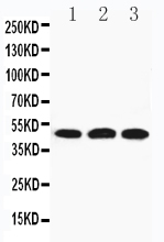
46.501 kDA

| Human | |
|---|---|
| Location: | 3p21.31 |
| Sequence: | 3; NC_000003.12 (50292832..50299405, complement) |
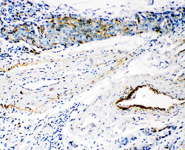
Expressed in sperm (PubMed:20586096). Highly expressed in epidermis of the skin, where it is expressed intracellularily in the deep horny layer (at protein level) (PubMed:21699545). Bone marrow, testis and kidney (PubMed:10493834).
Secreted. Cell membrane. Cytoplasmic vesicle, secretory vesicle, acrosome. Endoplasmic reticulum. Early endosome. Mostly present in low-density vesicles. Low levels in higher density vesicles of late endosomes and lysosomes. Localized in punctate cytoplasmic vesicles and in perinuclear structures, but does not colocalize with LAMP1. Localized on the plasma membrane over the acrosome and on the surface of the midpiece of the sperm tail.


PMID: 10493834 by Csoka A.B., et al. Expression analysis of six paralogous human hyaluronidase genes clustered on chromosomes 3p21 and 7q31.
PMID: 12084718 by Lokeshwar V.B., et al. Regulation of hyaluronidase activity by alternative mRNA splicing.