This website uses cookies to ensure you get the best experience on our website.
- Table of Contents
1 Citations 8 Q&As

Facts about Hyaluronidase-1.

.
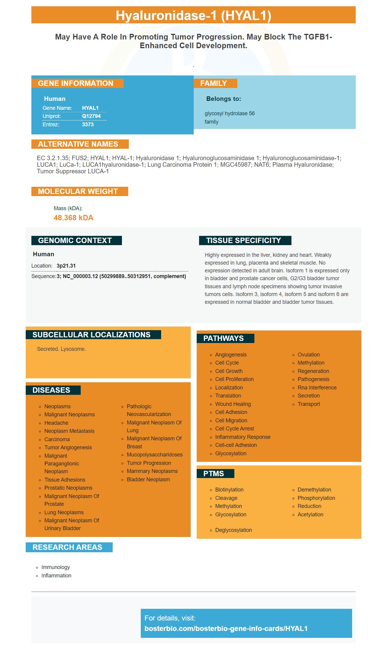
| Human | |
|---|---|
| Gene Name: | HYAL1 |
| Uniprot: | Q12794 |
| Entrez: | 3373 |

| Belongs to: |
|---|
| glycosyl hydrolase 56 family |

EC 3.2.1.35; FUS2; HYAL1; HYAL-1; Hyaluronidase 1; Hyaluronoglucosaminidase 1; hyaluronoglucosaminidase-1; LUCA1; luCa-1; LUCA1hyaluronidase-1; Lung carcinoma protein 1; MGC45987; NAT6; plasma hyaluronidase; tumor suppressor LUCA-1
Mass (kDA):
48.368 kDA

| Human | |
|---|---|
| Location: | 3p21.31 |
| Sequence: | 3; NC_000003.12 (50299889..50312951, complement) |
Highly expressed in the liver, kidney and heart. Weakly expressed in lung, placenta and skeletal muscle. No expression detected in adult brain. Isoform 1 is expressed only in bladder and prostate cancer cells, G2/G3 bladder tumor tissues and lymph node specimens showing tumor invasive tumors cells. Isoform 3, isoform 4, isoform 5 and isoform 6 are expressed in normal bladder and bladder tumor tissues.
Secreted. Lysosome.


PMID: 8603390 by Wei M.H., et al. Construction of a 600-kilobase cosmid clone contig and generation of a transcriptional map surrounding the lung cancer tumor suppressor gene (TSG) locus on human chromosome 3p21.3: progress toward the isolation of a lung cancer TSG.
PMID: 9223416 by Frost G.I., et al. Purification, cloning, and expression of human plasma hyaluronidase.
*More publications can be found for each product on its corresponding product page


