This website uses cookies to ensure you get the best experience on our website.
- Table of Contents
16 Q&As
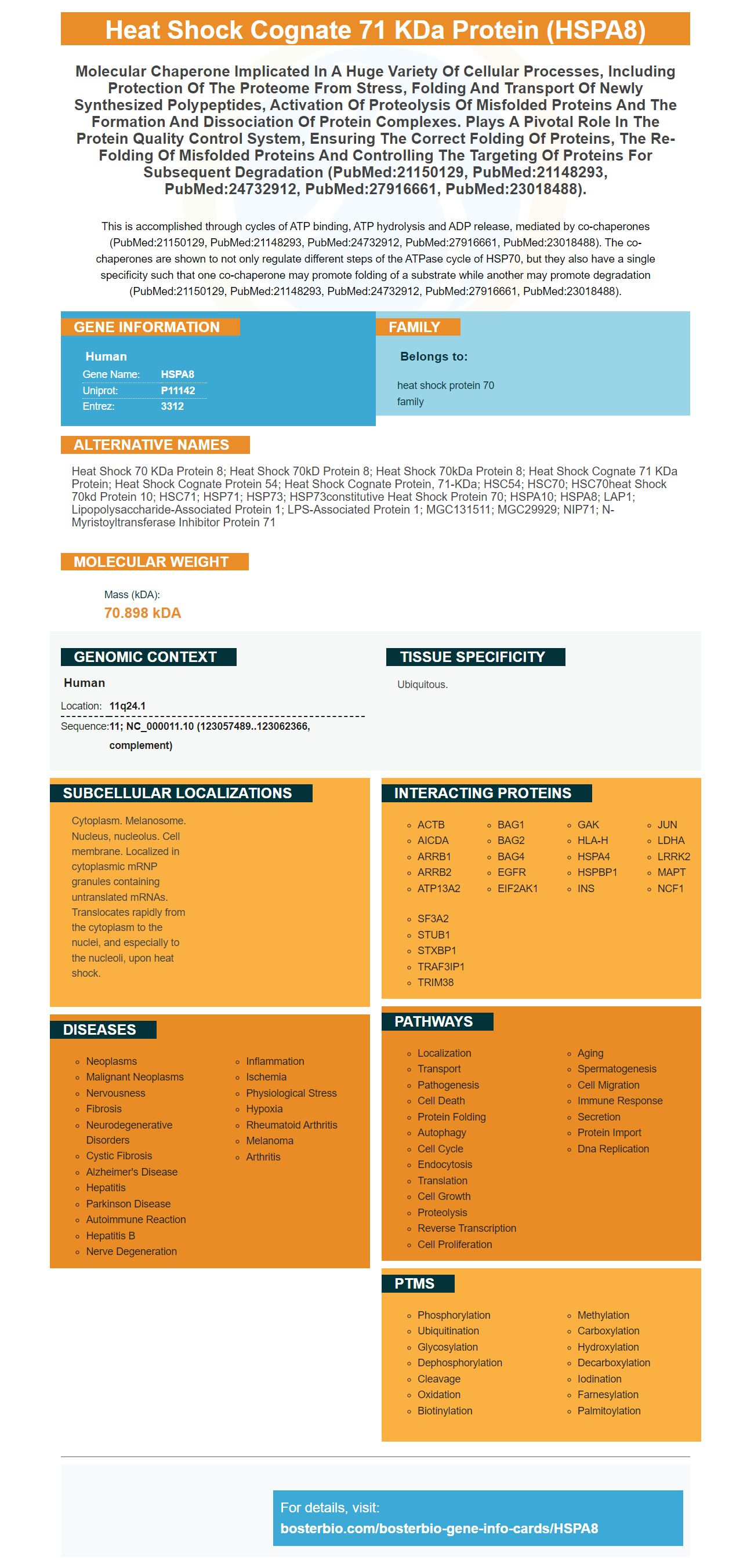
Facts about Heat shock cognate 71 kDa protein.
This is accomplished through cycles of ATP binding, ATP hydrolysis and ADP release, mediated by co-chaperones (PubMed:21150129, PubMed:21148293, PubMed:24732912, PubMed:27916661, PubMed:23018488). The co-chaperones are shown to not only regulate different steps of the ATPase cycle of HSP70, but they also have a single specificity such that one co-chaperone may promote folding of a substrate while another may promote degradation (PubMed:21150129, PubMed:21148293, PubMed:24732912, PubMed:27916661, PubMed:23018488).
Human | |
---|---|
Gene Name: | HSPA8 |
Uniprot: | P11142 |
Entrez: | 3312 |
Belongs to: |
---|
heat shock protein 70 family |
Heat shock 70 kDa protein 8; heat shock 70kD protein 8; heat shock 70kDa protein 8; heat shock cognate 71 kDa protein; heat shock cognate protein 54; heat shock cognate protein, 71-kDa; HSC54; HSC70; HSC70heat shock 70kd protein 10; HSC71; HSP71; HSP73; HSP73constitutive heat shock protein 70; HSPA10; HSPA8; LAP1; lipopolysaccharide-associated protein 1; LPS-associated protein 1; MGC131511; MGC29929; NIP71; N-myristoyltransferase inhibitor protein 71
Mass (kDA):
70.898 kDA
Human | |
---|---|
Location: | 11q24.1 |
Sequence: | 11; NC_000011.10 (123057489..123062366, complement) |
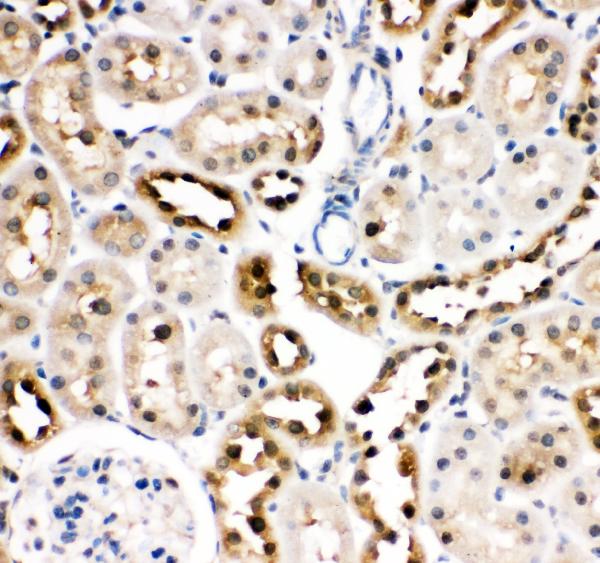
Ubiquitous.
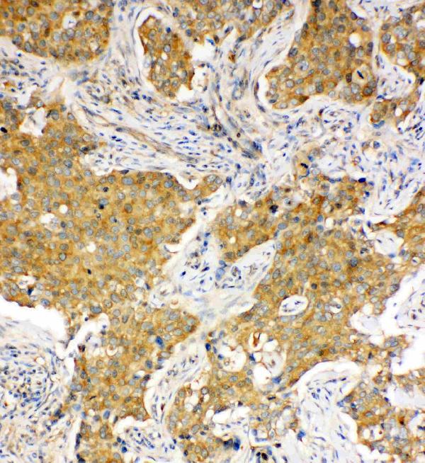
Cytoplasm. Melanosome. Nucleus, nucleolus. Cell membrane. Localized in cytoplasmic mRNP granules containing untranslated mRNAs. Translocates rapidly from the cytoplasm to the nuclei, and especially to the nucleoli, upon heat shock.
High-affinity antibodies for the HSPA8 marker are available in the market. The Boster Bio antibody validates the antibodies with known positive and negative samples to ensure high specificity and affinity. Boster Bio rewards the first reviewer of the product with credits and rewards scientists from all over the world. Read the article to learn more about these antibodies. Read the article to learn more about the Anti-Hsc70/HSPA8 Marker and its uses in molecular biology.
An Anti-Hsc70/HSPA6 or Anti-Hsc70/HSPA 8 marker has recently been introduced in the Boster Bio library, which identifies human Hsc70 protein. It is also a promising candidate for immunofluorescence studies. The antibody recognizes both human and rat Hsc70 protein. The antigen is available as Boster Bio product #BS488.
Interestingly, the antibody recognizes the Hsc70 protein, as well as other Hsc70 subunits. It is able to recognize these proteins by detecting their ubiquitin-modified lysine 10 residues. This protein is also a candidate for ALS. As its name implies, this antibody recognizes the Hsc70 protein found in the CNS.
This antibody was shown to decrease the level of APP in mouse models. It also reduced the levels of Ab plaque and APP protein. In addition, the Anti-Hsc70/HSPA8 Marker in Boster Bio blocked the effect of Metformin and inhibited the accumulation of APP in SH-SY5Y cells. However, Hsc70-S85A did not rescue the cytotoxic effect of overexpression of APP.
Hsc70 is a member of the HSP family. It has been found that it can dissociate conventional kinesin membrane vesicles. This protein is encoded by the ST13 gene and can interact with major cytosolic chaperones. However, it is absent from the mature receptor complex. Zhang and et al. (1998) mapped the ST13 gene to chromosome 22q13. The deletion of heterozygosity at 22q13 is common in colorectal cancer.
MRJ and HSC70 co-IP in Figure 1F are associated with a variety of complexes. This interaction could be caused by a direct association of HSC70 with IFT complexes. Interestingly, multiple antibodies against IFT88 and IFT57 co-precipitate the HSC70/HSPA8 marker. If HSC70/HSPA8 co-precipitates with the IFT88, this could suggest that anti-MRJ antibodies are also associated with this protein.
The Boster Anti-Hsc70/HSPA 8 Marker in Boster Bio is a versatile and sensitive antibody that enables you to perform a variety of immunological studies. The antibody is validated on multiple platforms and with known positive and negative samples. The peptide is highly specific and shows high affinity. Boster Bio also rewards first reviewers with product credits. The award is open to scientists around the world.
The HSPA8 protein is a constitutively expressed member of the heat shock protein (HSP) family, and it is abundantly expressed in neurons of mammalian brains. Recent studies show that constitutively expressed HSPA8 acts as a pre-protector for neurons against stress, possibly by targeting nuclear speckles with components of the protein disaggregation/refolding machine. Since HSPA8 is expressed in many neuronal cells, increasing its expression may be one additional way to combat protein misfolding.
The HSPA8 marker has been implicated in the regulation of many important biological processes, including the maintenance of cellular homeostasis. It participates in a wide range of cellular processes and provides important protection against environmental stresses. It is one of many members of the family of heat shock proteins (HSP) and exhibits diverse expression patterns, subcellular localizations, and functions. Heat shock proteins, HSP, interact with lipid membranes by interacting with phospholipid head-containing molecules. This interaction is facilitated by membrane insertion, and HSPA8 forms oligomerized complexes.
In addition to its role in refolding heat-damaged nucleolus proteins, HSPA1A and HSPA6 are also involved in the process of preventing aggregation. They are co-localized with HSPB1 and DNAJB1. The HSPA1A and HSPA6 protein markers, which co-localize with RNA splicing factors, were also identified in the same nuclei.
Liposomes contain different phospholipid moieties, and HSPA8 has a high propensity for insertion into these. HSPA8 binds to negatively charged phospholipids such as cardiolipin. The thermodynamic analysis of the interaction between HSPA8 and liposomes showed that the membrane insertion occurs spontaneously, but nucleotides blocked this interaction.
Furthermore, HSPA8 is an important factor in the regulation of neuronal cell differentiation. Its targeting properties are similar to those of HSPA1A, but different from those of HSPA6. HSPA8 is localized at nuclear speckles with machine components and is targeted to the GC layer of the nucleus along with BAG-1. This data provides new insights into neuronal development.
High-affinity primary antibodies using the HSP8 marker from Boster Bio have been validated in multiple platforms and are tested against known positive and negative samples. Boster also rewards the first reviewer of its products with product credits and provides a free blocking peptide for the specific immunogen length. Scientists worldwide are encouraged to review Boster's high-affinity primary antibodies.
The process for making polyclonal primary antibodies is based on the immune systems of hosts. It is made possible by using proteins called complementarity-determining regions, which are the building blocks of antibodies. Complementarity-determining regions are a complicated topic. You can learn more about them on Wikipedia. In the process of creating polyclonal antibodies, scientists first immunize host animals with antigens and then isolate antibodies from their sera and eggs.
Another advantage of this method is its dual labeling capability. This means you can use one primary antibody to label specimens with a secondary antibody and vice versa. This allows you to ask more questions with a single specimen, giving you a stronger answer and broader contextual data. And because of the versatility of the Boster Bio High-affinity primary antibodies using the HSPA8 marker, you can perform multiple experiments at the same time.
The current phase III clinical trial of HSPA8 targets this gene, which acts as a chaperone for class II major histocompatibility complex proteins. Alterations in the trafficking of this gene may have implications for the presentation of antigenic peptides to T cells and their downstream immune response. Several validation methods were used to determine whether HSPA8 is associated with this type of cancer. The study results indicate that there are several important pathways involved in the process of tumor development, such as HSPA8.
The predictive capacity of HSPA8 was similar to other known prognostic factors, including age and gene mutations. However, its predictive capacity for karyotype abnormalities was lower. The findings suggest that HSPA8 is a viable candidate marker for cancer treatment. However, further research is needed to confirm the results of these studies. The validation methods for HSPA8 Marker are discussed below. Once these methods are validated, the HSPA8 Marker should be widely used in clinical trials of karyotype abnormalities.
The cytoplasmic HSPA8 is present in cells under normal conditions, but it is transiently exported in the cytoplasm during recovery. Upon oxidative stress, it is required for cell survival. However, during heat stress, HSPA8 is imported into the nucleus and egresses between three and six hours. The abundance of HSPA8 was expected due to its presence in the cytoplasm but was not detected. In addition, HSPA8 content in the nucleus was undetectable in total cells.
To validate HSPA8 Marker, two approaches were used: proteomics analysis and crosslinking experiments. These methods revealed that HSPA8 binds to the P140 molecule. The P140 effect is believed to result from binding to the nuclear import signal sequences, which are included in the HSPA8 structure. Further, the P140 peptide is associated with the expression of tumor suppressors in the nucleus.
PMID: 3037489 by Dworniczak B.P., et al. Structure and expression of a human gene coding for a 71 kd heat shock 'cognate' protein.
PMID: 11093761 by Tsukahara F., et al. Molecular and functional characterization of HSC54, a novel variant of human heat shock cognate protein 70.