This website uses cookies to ensure you get the best experience on our website.
- Table of Contents
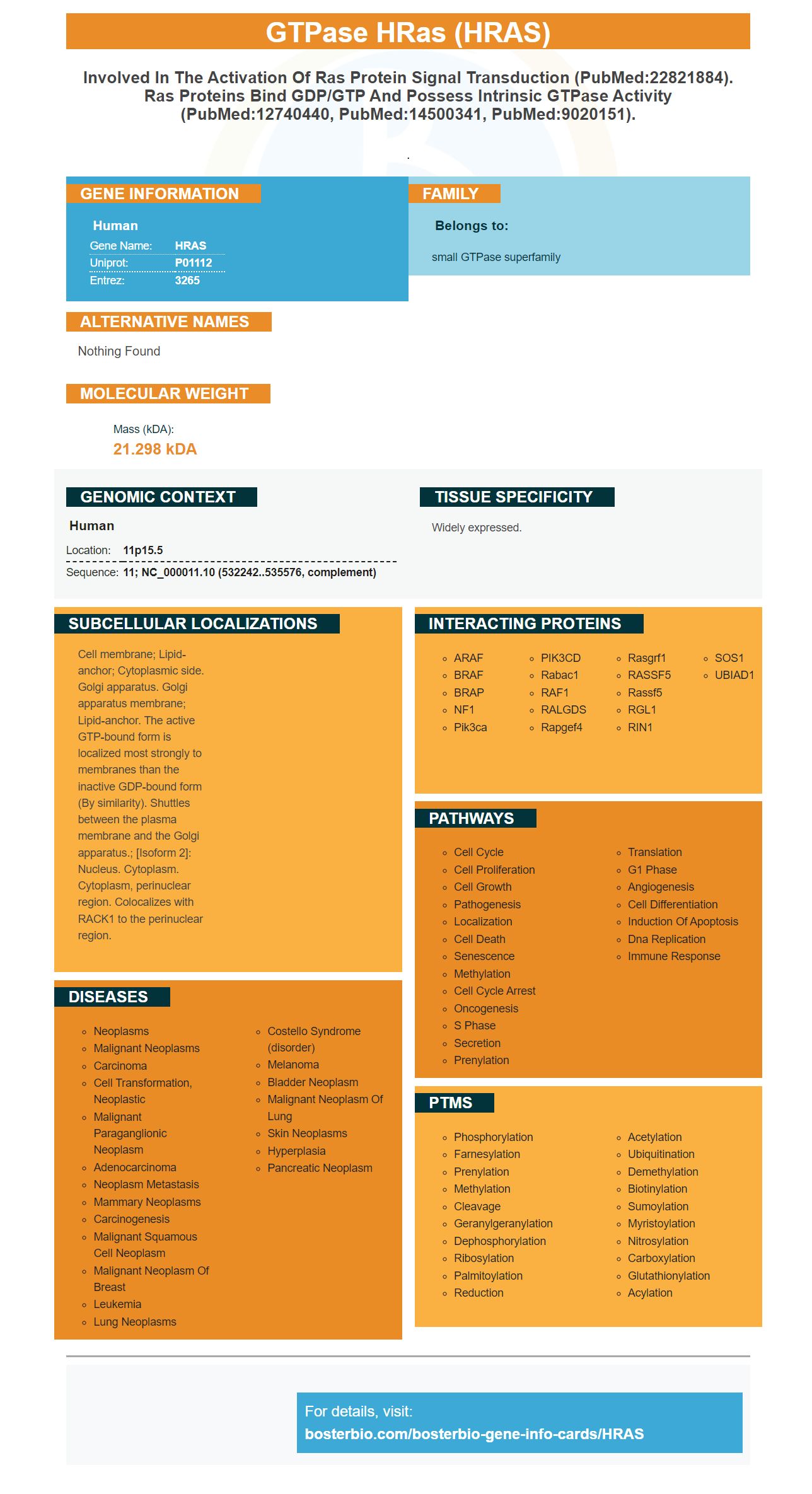
Facts about GTPase HRas.
.
Human | |
---|---|
Gene Name: | HRAS |
Uniprot: | P01112 |
Entrez: | 3265 |
Belongs to: |
---|
small GTPase superfamily |
Nothing Found
Mass (kDA):
21.298 kDA
Human | |
---|---|
Location: | 11p15.5 |
Sequence: | 11; NC_000011.10 (532242..535576, complement) |
Widely expressed.
Cell membrane; Lipid-anchor; Cytoplasmic side. Golgi apparatus. Golgi apparatus membrane; Lipid-anchor. The active GTP-bound form is localized most strongly to membranes than the inactive GDP-bound form (By similarity). Shuttles between the plasma membrane and the Golgi apparatus.; [Isoform 2]: Nucleus. Cytoplasm. Cytoplasm, perinuclear region. Colocalizes with RACK1 to the perinuclear region.
This article will talk about how to use Boster Bio's Antibodies and the PCR conditions for detecting HRAS protein. Boster Bio's antibodies are available through various vendors, including IDT and Life Technologies. This article will be most useful to people who are new to molecular biology and would like to learn more about how to use antibodies. Also, it will give some useful tips for using Boster Bio's products.
The HRAS marker can be used to identify different types of antibodies. This marker has multiple applications, including the production of polyclonal antibodies and monoclonal antibodies against the capsular polysaccharides of Neisseria meningitidis. This marker can also be used for the preparation of latex reagents. Purchasing antibodies from Boster Bio is a smart way to ensure that you receive high-quality and consistent antibodies.
PCR conditions used for the detection of HRAS markers were the same as those for the analysis of KRAS gene. The primers were CCGGAAGGTCCTGAGGGG and CACGGAAGGTCCTGAGGGG respectively. The PCR reaction volume was 20 mL. One ml of template was added to the reaction mixture. The primers are given in the Supplementary Material.
The HRAS gene belongs to a group of genes known as oncogenes. Oncogenes are responsible for causing cancer in normal cells. HRAS is part of the Ras family, which also includes KRAS and NRAS. The Ras family produces GTPases that play important roles in cell division, differentiation, and apoptosis. A high-quality HRAS marker will show whether the DNA of a patient contains the gene HRAS.
In this case, the Cre promoter is expressed in an auto-excision construct. The resultant PCR product will be a HRAS marker. The RNA from this reaction will be a DNA fragment containing the coding sequence of the corresponding HRAS gene. The PCR product will include the primers and the probe. The primers should be designed such that each strand is unique. The primers should be spaced one millimeter apart.
There are several types of mutations in the HRAS gene, which can derail the response of a patient to targeted therapies. However, little is known about the frequency of these mutations in breast cancer. For instance, only nine out of nine DCs contained detectable levels of at least one of the five mutations that were observed in DCs. In addition, HRAS G12D was elevated significantly in DCs.
The HRAS and KRAS genes were expressed in the same way in the blood samples obtained from the same patient at three observation points. For each sample, tissue samples and blood samples were collected before and after surgery. This allowed the researchers to obtain accurate quantitative values from the HRAS marker. This study showed that the HRAS gene was highly expressed in cancer patients with a tumor that was sized at least 50 mm in length.
The ACB-PCR method detects initiating mutations of the H-ras gene in 15 out of 21 tumors. However, the results of the mutant fractions are not reliable because the sample could contain contaminating normal tissues. To make sure, ACB-PCR is performed under optimal conditions to confirm the presence of HRAS mutations in tumors. Therefore, it is important to perform a multicenter study to confirm the findings.
Detection of HRAS protein by boster bio uses a polyclonal antibody. This antibody reacts with the GTPase HRAS in many animal samples. Boster Bio develops antibodies for a variety of samples from rabbits and mice. The active GTP-bound form of the protein localizes to membranes, while the inactive GDP-bound form is largely distributed throughout the cell. The proteins are shuttled between the Golgi apparatus and plasma membrane.
PMID: 6298635 by Capon D.J., et al. Complete nucleotide sequences of the T24 human bladder carcinoma oncogene and its normal homologue.
PMID: 6844927 by Reddy E.P.; Nucleotide sequence analysis of the T24 human bladder carcinoma oncogene.