This website uses cookies to ensure you get the best experience on our website.
- Table of Contents
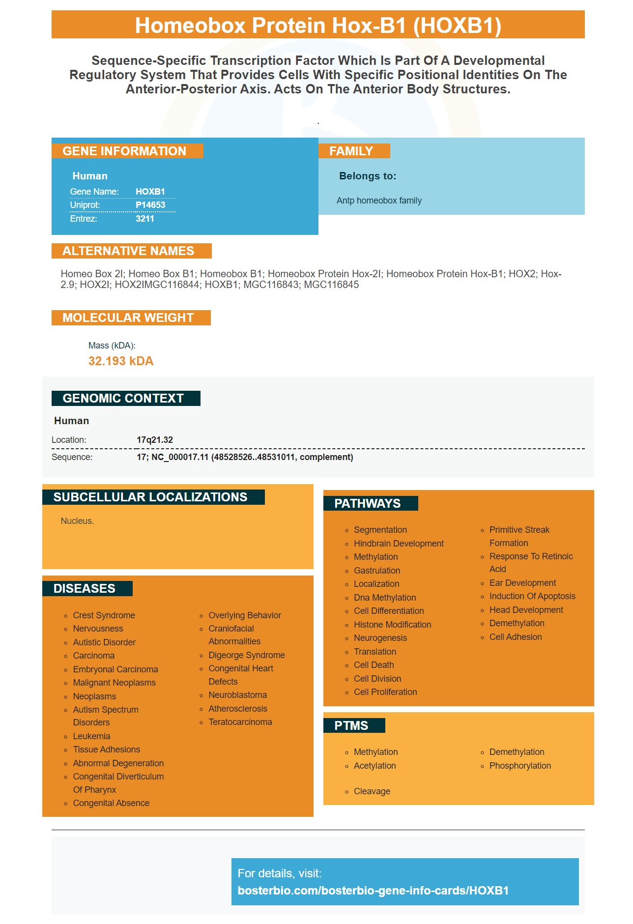
Facts about Homeobox protein Hox-B1.
.
Human | |
---|---|
Gene Name: | HOXB1 |
Uniprot: | P14653 |
Entrez: | 3211 |
Belongs to: |
---|
Antp homeobox family |
homeo box 2I; homeo box B1; homeobox B1; Homeobox protein Hox-2I; homeobox protein Hox-B1; HOX2; Hox-2.9; HOX2I; HOX2IMGC116844; HOXB1; MGC116843; MGC116845
Mass (kDA):
32.193 kDA
Human | |
---|---|
Location: | 17q21.32 |
Sequence: | 17; NC_000017.11 (48528526..48531011, complement) |
Nucleus.
The HOXB1 marker is a very popular gene for research on human reproduction. It co-operates with MEIS1 to regulate embryonic uterine development. Here's a brief review of this gene's best uses. This article was written for scientists who are interested in furthering their knowledge of human reproductive health. Read on to learn more about this gene and how it can be used to improve your reproductive health.
HOXA10 is a homeobox transcriptional factor that regulates several receptive-related genes. The transcriptional activity of HoxA10 is required for decidualization, which creates the microenvironment for implantation. Loss of HoxA10 causes failed implantation during in vitro fertilization treatments. Loss of HoxA10 impairs stromal cell proliferation during decidualization and arrests the cell cycle.
There are several biomarkers of endometrial receptivity. MicroRNA-135b is related to endometrial receptivity, including Circ-8073, CEP55, and av b3. Both markers are associated with the endometrium's receptivity. In addition, Circ-8073 has been implicated in endometrial receptivity development in dairy goats.
IL-10 is a known regulator of HOXA10, a receptivity marker. In a study, the level of HOXA10 and IL-10 was decreased in adenomyosis-associated women. Further investigation is needed to determine whether IL-10 plays a role in endometrial receptivity and embryo implantation.
HOXA10 is a marker that shows whether the endometrium is receptive to hCG. The age of the woman's uterus and HOXA10 expression have been linked to the receptivity of the endometrium. In the study, LIF were the markers that were most highly expressed. LIF and HOXA10 increased significantly in the MH group. This study also demonstrates that uterine age and HOXA10 are important indicators for detecting implantation.
Although endometrial receptivity can be assessed by a variety of methods, HOXA10 has recently been applied in a clinical trial at the Malhotra Test Tube Baby Centre. This method, which involves the use of customised microarrays for genes involved in endometrial receptivity, can predict endometrial dating. The genetic profile of the endometrium compared with LH + 7 and P + 5, a natural hormonal cycle, predicts the window for implantation.
This study suggests that metformin may improve endometrial receptivity in women with PCOS. However, the exact mechanism by which metformin improves endometrial receptivity is not fully understood. This study shows that metformin treatment increases the expression of HOXA10 and ITGB3 in the endometrium of PCOS women.
In a study of female Wistar albino mature rats, the effects of metformin on endometrial receptivity were determined. The study also examined the effects of metformin on the expression of LIF and HOXA10. In addition to endometrial receptivity, it demonstrated that metformin treatment reduced the expression of MMP-9 and VEGF in mice. This study identifies a molecular pathway for endometrial receptivity and fertility.
In another study, miR-135b inhibits the expression of HOXA10 by lowering the luciferase activity of HoxA10-WT and HoxA10-MUT cells. However, hsa_circ_001946 reverses this effect and increases HOXA10 protein expression. Interestingly, these results are in line with previous studies.
The co-operating gene MEIS1 was discovered through the work of Hoxb1 and its partner HOXB1. The transcriptionally activated form of MEIS1 is called E2A-PBX1. The results showed that Vp16-Meis1 induces basal transcription of its signature genes. In contrast, Vp16-Meis1 had no effect on NIH 3T3 fibroblasts.
Meis1 regulates the expansion of leukemic stem cells and is required for their self-renewal. In addition, Hoxa7 and Hoxa9 enhance the expression of Meis1-related signature genes in Vp16-Meis1 progenitors. This interaction leads to the activation of Meis1-related genes by Hox proteins in normal hematopoiesis.
Hoxa9 is necessary for the immortalization of FL-dependent and SCF-dependent progenitors. It also mediates the production of Flt3 in ScaI-Meis1 progenitors. Hoxb1 is dispensable for the upregulation and proliferation of Flt3 in Vp16-Meis1 progenitors. Mutations in Hoxb1 prevent this interaction.
The study demonstrates that HOXB1 is a cooperating gene with MEIS1. However, the function of Hoxa9 is critical for the expression of signature genes. In addition, Meis1 interacts with a transcription factor called Pbx3. Hoxa9 and Pbx3 act as a co-operating complex, which is crucial for the activation of Hoxa9 and Meis1 genes.
Meis1 and Pbx3 co-expressed in Cells 24 h after transfection. Flag-tagged protein was detected by immunoblotting. B-actin served as the loading control. Its expression has been implicated in leukemogenic activity. Despite its intrinsic stability, MeisD has no function in CFC assays.
Both Meis1 and PREP1 play important roles in myeloid leukemia. However, MEIS1 and PREP1 share some functional similarities in the activation of Hox/Meis1 signature genes. This is important for further research on the genetics of leukemia. The two co-operating genes have several distinct roles in leukemia, but they are still a common collaborator.
Mutations in HOXB1 disrupt the protein's DNA-protein interactions. The resulting disruptions in the protein's activity may affect the cell's response to hormones. Therefore, a mutation in HOXb1 may have a profound impact on the cell's development. Hoxb1 co-operates with MEIS1 in the regulation of cell growth.
The structural and functional properties of Meis1 are conserved across different organ and cellular systems. Its function in diverse organ systems suggests that it is important for combinatorial diversity and gene spectrum. If Meis1 is essential for the development of human embryos, HOXB1 is a key player in the process. But what does this mean for MEIS1's stability?
HOXB1 and MEIS1 share several functional properties. Both MeisD and HOXB1 have a transcriptional function. Their co-operating activity was also confirmed by a graphical representation of the interaction. Hoxa9DPbx activity is compromised by the loss of Pbx interactions with Meis1.
Hoxa proteins, including HOXB1, occupy critical roles in the patterning of embryonic uterine development and have also been implicated in the development of endometrium and the implantation of the embryo. HOXA10 and HOXB1 are expressed in endometrial glands and stroma during the menstrual cycle, and are essential for embryo implantation in both humans and mice. Mutations in either gene result in impaired endometrial receptivity, resulting in female infertility.
The activity of the gene is controlled by a transmembrane protein, called the Gal4 polypeptide. This protein spreads transcriptional activity over its entire sequence and binds multiple ATTA-containing sites in the HCR. It also binds to the Gal4-responsive element. It also has a C terminus containing 71 residues, which is highly conserved among group 3 Hox proteins.
The genes within the HOXA cluster lack the homologue of the corresponding gene in humans. HOXA1 and HOXB1 are highly conserved across species and develop characteristic spatial distribution throughout the Mullerian duct. Defects in these genes cause region-specific defects along the female reproductive tract. For example, deficiency of Hoxa10 results in homeotic transformation of the anterior uterus. In addition, Hoxa10 and Hoxa13 null embryos exhibit hypoplastic urogenital genital sinuses and agenesis of the posterior part of the Mullerian duct.
The expression of Hox genes is compared between different stages of embryonic uterine development. Hoxb1 is expressed at the sinus venosus while Hoxa2, c, and d are expressed at later stages of development. Furthermore, miR-130a and miR-106a have significant effects on the expression of the Hox genes. The H9c cardiomyoblasts have a low expression of Hox genes.
In addition, circulating factors such as cytokines and chemokines have important roles in embryo-mother cross communication. EVs are important during preconception, implantation, and pregnancy, and the presence of EVs in the uterus has only recently become a topic of interest. But since they have specific roles in embryo-maternal communication, they may play key roles in preconception, pregnancy, and implantation. The first evidences for EVs in human uterine fluid were presented by Ng et al. and Burns et al. Eventually, these EVs were confirmed to contain exosomal markers and have an essential role in embryonic development.
PMID: 2574852 by Acampora D., et al. The human HOX gene family.
PMID: 2576652 by Boncinelli E., et al. Organization of human class I homeobox genes.