This website uses cookies to ensure you get the best experience on our website.
- Table of Contents
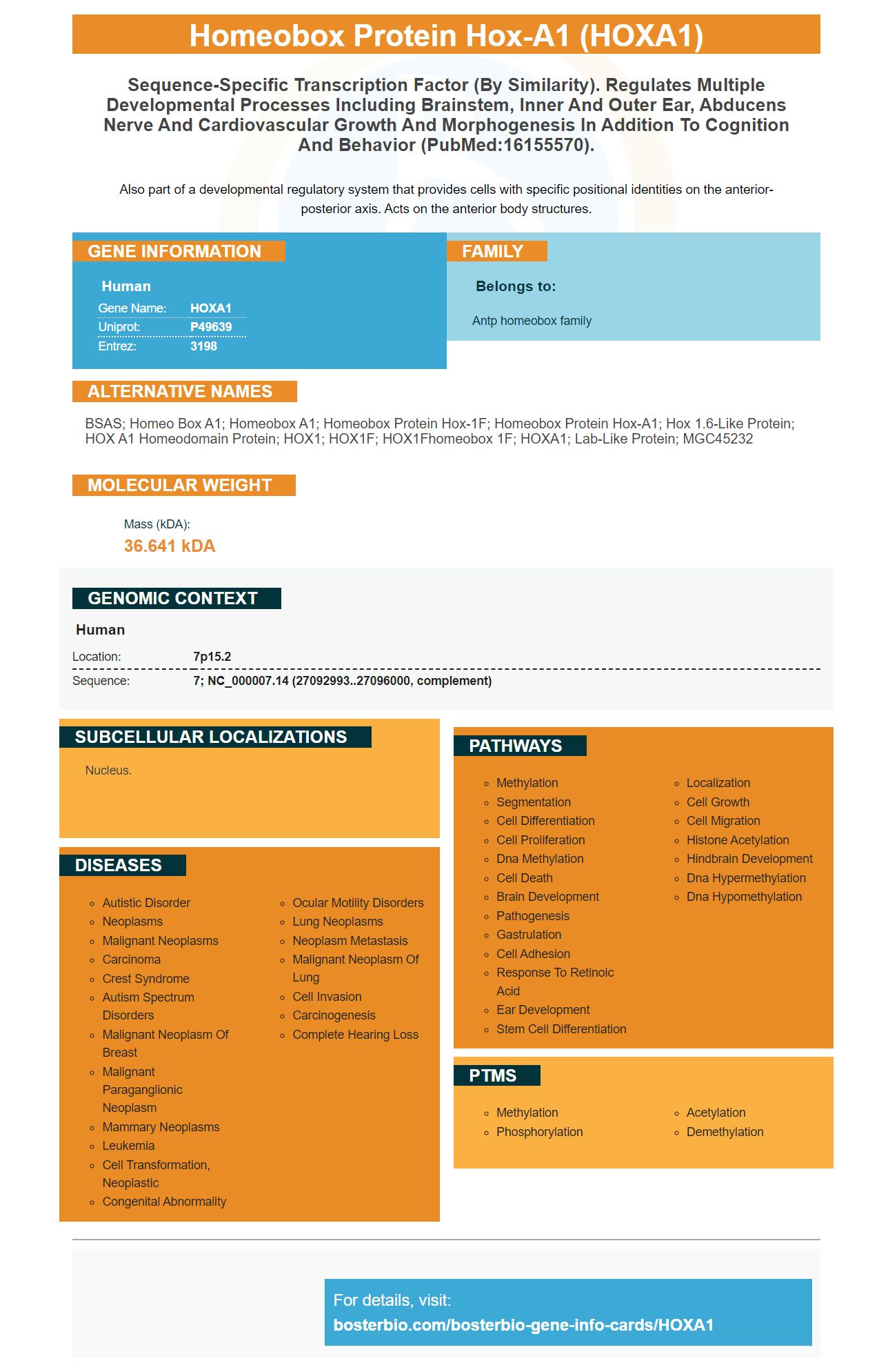
Facts about Homeobox protein Hox-A1.
Also part of a developmental regulatory system that provides cells with specific positional identities on the anterior-posterior axis. Acts on the anterior body structures.
Human | |
---|---|
Gene Name: | HOXA1 |
Uniprot: | P49639 |
Entrez: | 3198 |
Belongs to: |
---|
Antp homeobox family |
BSAS; homeo box A1; homeobox A1; Homeobox protein Hox-1F; homeobox protein Hox-A1; Hox 1.6-like protein; HOX A1 homeodomain protein; HOX1; HOX1F; HOX1Fhomeobox 1F; HOXA1; lab-like protein; MGC45232
Mass (kDA):
36.641 kDA
Human | |
---|---|
Location: | 7p15.2 |
Sequence: | 7; NC_000007.14 (27092993..27096000, complement) |
Nucleus.
Steven Boster invented his first product in 1993. He earned the nickname "he that converts science into the lavatory" Boster has been creating primary antibodies for hundreds of years and has become one of China's largest catalog antibody companies. PicoKine(tm), Boster's own proprietary ELISA platform was launched in the late nineties. This system uses proprietary trade secrets in order to create high-sensitivity ELISA kit.
The HOXA1 genome is a vital candidate for recombinantDNA tech and has many uses, including in vitro diagnosing. The SUMO1–HOXA10 marker was used during timed mid-secretory entometrial biopsies. This study showed that the gene was abnormally expressed in RIF and controls, and that abnormal SUMO1 sumoylation could contribute to impaired endometrial receptivity or implantation in RIF women.
HOXA10 not only plays a role in oocyte production, but it is also an important molecular marker to determine endometrial receptivity. Recurrent failures in implantation have been linked to impaired endometrial responsiveness. SUMO1 binds HOXA10 and inhibits its stability, transcriptional activity, and mRNA expression. HOXA10 is also associated with adenomyosis, hydrosalpinges, and infertility.
SUMO1 ubiquitination can also be affected by HOXA1. Flag-SUMO1 overexpression inhibited HOXA10-mediated attachment, but did not impair the SUMO1-promoted marker's ability to promote cell adhesion. Moreover, SUMO1-promoted attachement was not affected by overexpression HOXA10K164R.
The HOXA1 gene, which is essential for embryo implantation, has many applications. The HOXA10 family includes a number of transcription factors, many of which have high-conserved homeodomains and high DNA-binding activity. It has been implicated as a factor in tumorigenesis, fibroblast differentiation, and other processes. HOXA10 has been implicated in tumor and cancer research. It is useful for identifying ovarian tumour cells and determining the effect of SUMO conjugate on human embryos.
The differential regulation by Hoxa1 has implications on a number physiological processes, such as neural crest growth, membrane dynamics, vesicular transportation, and membrane dynamics. BiFC assay demonstrated interactions between Hoxa1 & different signaling pathways such as focal adhesion and axon-guidance. These proteins, however, are not directly involved in the regulation of gene expression and may have indirect effects on the biology of a cell.
The bimolecular fluorescence complementation assay (BiFC) assay was developed to visualize distinct interactions between Hoxa1 and its partners in live cells. Using the fusion protein Venus, the ORF of 45 validated Hoxa1 interacting proteins was fused to the C-terminal moiety of a fluorescent protein. The BiFC assay revealed a specific interaction of Hoxa1 with PBX1A.
Transfection was achieved using MATaY8930, MATaY8800. Then, NaCl 120mM, EDTA 0.5mM, NP40 0.5%, and glycerol 10% were added to the culture. To aid in the growth of the clone, cells were also supplemented with tryptophan and leucine.
SP140 is an epigenetic “reader” that recognizes the H3K27me3 & H3K27ac marks in histones. It preferentially interacts in macrophages with chromatin in TSSs containing genes expressing the repressive H3K27me3 and H3K27ac. Also, SP140 is positively correlated to H3K27me3 enrichment near enhancer areas. Depletion of SP140 can lead to impaired innate immunity. This is important for maintaining intestinal homeostasis.
GATA6R456G/R456G cellular epigenetic profiles are possible due to aberrant signaling by RA. GATA6+/R456G cellular transcript levels of NR2F2, ZFPM2, HOXA1 were higher than WT on differentiation day 4. It has been demonstrated that HOXA1 haploinsufficiency can interfere with the regulation and expression of many genes during neural differentiation.
The results from the NANOG binding genomic region showed that Pou3f1 was regulated by HOXA1 & NANOG. This genomic region is deleted, which results in increased Pou3f1 transcription. The NANOG binding genomic region's Pou3f1 gene expression is indicated by the dashed lines. Therefore, the results from the NANOG-bound genomic region are highly relevant to our understanding of embryonic development.
HOXA1 is a member of a family proteins whose gene products are vital for the development and maintenance of human tissues. This protein performs multiple functions. It also directs the expression genes that are crucial for the specification and development of neural crests. It is a critical component of vertebrate morphogenesis, as evidenced by its required role in the morphogenesis of external genitals and digits. Numerous studies have identified HOX genes in morphogenesis.
Tischfield et.al. discovered the gene HOXA1. by homologous recombination. This mutation leads to homozygosity of a transition between a stop codon and an arginine amino acid. This mutation does not exist in the control DNA, which supports the founder effect theory. However, functional analysis in patient cells has not been included in these studies.
HOXA1 is a protein that regulates signaling pathways within ESCs that control differentiation. Hoxa1 either promotes or inhibits the expression target genes. Genes targeted by Hoxa1 include Cdx1, Snai1, Klf2, and Sall1.
CLNSS cells had significantly increased expression of HOXA1 with both hPSCs (and CLNSCs), and this was due to a region-specific pattern. RNA-seq analyses of the four genes showed significant enrichment for spinal cord-related GO functional terms. CLNSCs expressed more HOXA1 than any other gene, which confirms their neural phenotype.
The development and maintenance of digits and genitals, both external and inner, in vertebrates depends on the transcriptional regulation of HOXA1. The shared enhancer-containing landscape of HOXA1 and HOXD1 genes was studied. They share a regulatory spine, and tissue-specific interactions suggest hijacking of this regulatory spine. It was also found that HOXA1 & HOXD1 were distinct by genome duplication in ancestral animals. Moreover, it was suggested that preexisting chromatin architecture facilitates recruitment of transcription factors.
A full-length CDS of HOXA1 was cloned into the pLCH72 vector to test HOXA1 gene transcription in CLNSCs. After transfection, ESCs received puromycin treatment and cell lines were analyzed. The cells that were transfected successfully were then examined by western blotting or qRTPCR.
Autism susceptibility may also include HOXA1 gene transcription. A genetic variation within rhombomeres 4-6 could also contribute. Common polymorphisms in rhombomeres 4-7 have been linked to an increase in autism risk in humans. Knockout mice with a rhombomere 4-7 gene mutation exhibit altered embryonic development of facial nuclei, superior olivary nuclei, and ear shape.
Despite the fact HOXA1 being inactive in healthy oral tissues it has been linked to cancer. HOXA1 gene expression has been linked to tumorigenesis and can be used in OSCC as a prognostic tool. It is important that HOXA1 expression is higher in OSCC than in healthy oral tissues. Patients with cancer of their mouths are at higher risk for developing oral-related complications than patients with healthy mucosa.
PMID: 7622051 by Hong Y.S., et al. Structure and function of the HOX A1 human homeobox gene cDNA.
PMID: 7488013 by Chariot A., et al. Retinoic acid induces three newly cloned HOXA1 transcripts in MCF7 breast cancer cells.