This website uses cookies to ensure you get the best experience on our website.
- Table of Contents
21 Q&As
Facts about HLA class I histocompatibility antigen, C alpha chain.
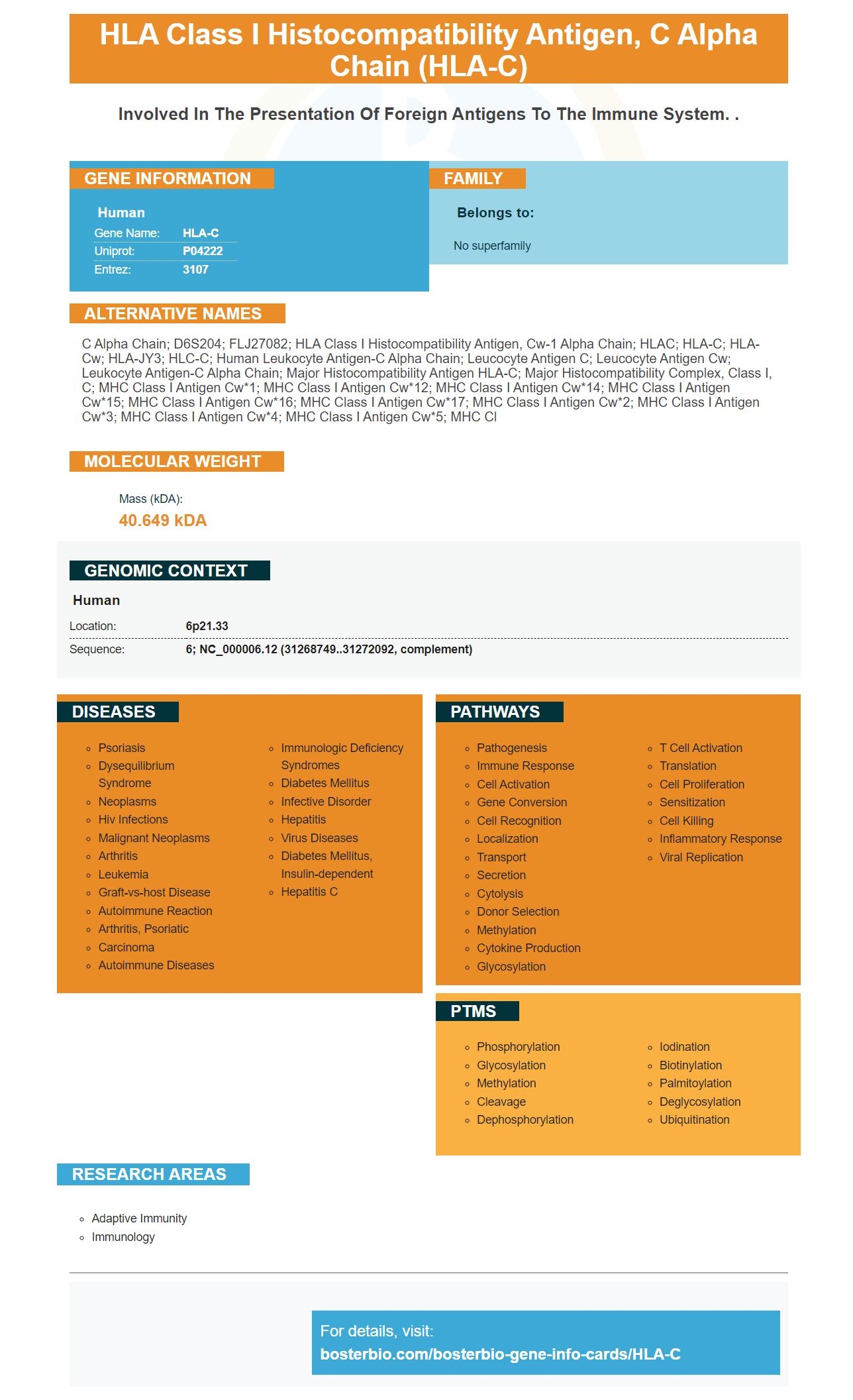
Human | |
---|---|
Gene Name: | HLA-C |
Uniprot: | P04222 |
Entrez: | 3107 |
Belongs to: |
---|
No superfamily |
C alpha chain; D6S204; FLJ27082; HLA class I histocompatibility antigen, Cw-1 alpha chain; HLAC; HLA-C; HLA-Cw; HLA-JY3; HLC-C; human leukocyte antigen-C alpha chain; leucocyte antigen C; leucocyte antigen Cw; leukocyte antigen-C alpha chain; major histocompatibility antigen HLA-C; major histocompatibility complex, class I, C; MHC class I antigen Cw*1; MHC class I antigen Cw*12; MHC class I antigen Cw*14; MHC class I antigen Cw*15; MHC class I antigen Cw*16; MHC class I antigen Cw*17; MHC class I antigen Cw*2; MHC class I antigen Cw*3; MHC class I antigen Cw*4; MHC class I antigen Cw*5; MHC cl
Mass (kDA):
40.649 kDA
Human | |
---|---|
Location: | 6p21.33 |
Sequence: | 6; NC_000006.12 (31268749..31272092, complement) |
In this article, we'll cover the Boster Bio Anti-HLA-C Marker and its use in IHC workflows. In addition, we'll discuss the Boster Bio IHC protocol. This is a vital part of using this antibody. It can help you make the most of your Boster Bio kit. Read on for more information. But before we dive in, let's review the Boster Bio workflow.
The HLA-C marker is a widely used biomarker for identifying certain types of cells in the body. Boster Bio has developed a proprietary ELISA platform that allows researchers to use the HLA-C gene to detect and analyze human immune cells. The ELISA platform is proprietary and uses trade secrets to produce highly sensitive ELISA kits. Boster Bio also offers a comprehensive library of antibodies and other diagnostic tools for the HLA-C Marker.
The classical HLA loci are extremely diverse and close to several genes driving innate immunity. These loci are difficult to dissect genetically, but the disease has a long history of genetic studies and large cohorts, which provides statistical power for studying the effects of specific HLA alleles. Boster Bio: Best Uses Of The HLA-C Marker
Reverse immunogenetics is a natural extension of HLA-infection associations. However, the resulting failure of the approach to produce a clinically effective vaccine highlights the complexity of genetic variation in pathogens. Vaccines based on HLA-C restriction may not reach a large proportion of the population at risk. That is why it is critical to study the HLA-C gene and its associations with human immunity.
To detect antibodies against HLA-C, tissue sections were incubated with a mix of AP-labelled primary anti-mouse antibodies and HRP-labelled secondary anti-rabbit antibodies. The tissue sections were developed with a Vector Blue Substrate kit. After staining, the sections were dehydrated and mounted using Ecomount (Biocare Medical).
The sample was cut into 4mm sections and rehydrated. Sections were then heat-mediated antigen retrieval in EnvisionTM FLEX target retrieval solution (DAKO). After immunohistochemical staining, the slides were counterstained with the appropriate secondary antibodies. The immunohistochemical reaction was performed on each slide in duplicate. The results were compared with each other and with the reference standard.
The IHC protocol for the HLA-C mark uses a horseradish peroxidase system or an alkaline phosphatase-based staining procedure. Both of these methods require that endogenous enzymes be blocked to avoid non-specific binding. The resulting non-specific binding of the secondary antibody can occur due to residual sites on the tissue section. Blocking should also be done carefully as serum from the same species may bind to sites prior to IHC and result in false positive results. Blocking should be limited to 10-30 minutes and should not be overly intensive.
To perform the IHC protocol for the HLA-C mark, tissue samples must be prepared. Tissues can be obtained from biopsy, animal model, or surgery. Surgical tissue samples are obtained after the animal has died for at least 2 hours. The autopsy specimen is similar to the postmortem autolysis process, but the antigens may denature. Once the animal has died, the tissues should be fixed to ensure that the test is accurate and reproducible.
The performance of anti-CD163 antibody was best after overnight incubation at 4degC. This protocol is the same for HLA-B27, but anti-CD163 showed better results after overnight incubation at 4degC. Once the cytology results are obtained, the anti-CD163 antibody must be diluted and incubated for 5 more hours. Then the IHC protocol for the HLA-C marker should be completed.
This article describes a Boster Bio workflow for HLA-C testing. The Picoband reagent reacts with Human, Mouse, and Rat cells and is available for flow cytometry. It contains 5 mg of BSA. For immunofluorescence analysis, the Picoband contains anti-HLA-C antibody. Boster Bio's workflow for HLA-C testing was developed by Dr. John Borland and colleagues.
If you're in need of a quality anti-HLA-C marker, Boster Bio can help. This company specializes in antibody manufacturing, including ELISA and signature products. More recently, they've also started offering PCR-related molecular biology products. In addition to offering a wide range of products, Boster Bio also provides various services for scientists, such as free technical resources and 24 hour support.
Boster Bio Anti-HLA-C Antibody Picoband is tested in flow cytometry, ICC, and IHC-P. It reacts with Human, Mouse, and Rat HLA-C molecules. It contains five milligrams of BSA and is suitable for use in flow cytometry. This product also reacts with several other HLA-C markers. It is highly recommended for use in studies in which you'd like to examine HLA-C levels in your samples.
PMID: 2914713 by Cianetti L., et al. Three new class I HLA alleles: structure of mRNAs and alternative mechanisms of processing.
PMID: 1711567 by Grassi F., et al. Human immunodeficiency virus type 1 gp120 mimics a hidden monomorphic epitope borne by class I major histocompatibility complex heavy chains.