This website uses cookies to ensure you get the best experience on our website.
- Table of Contents
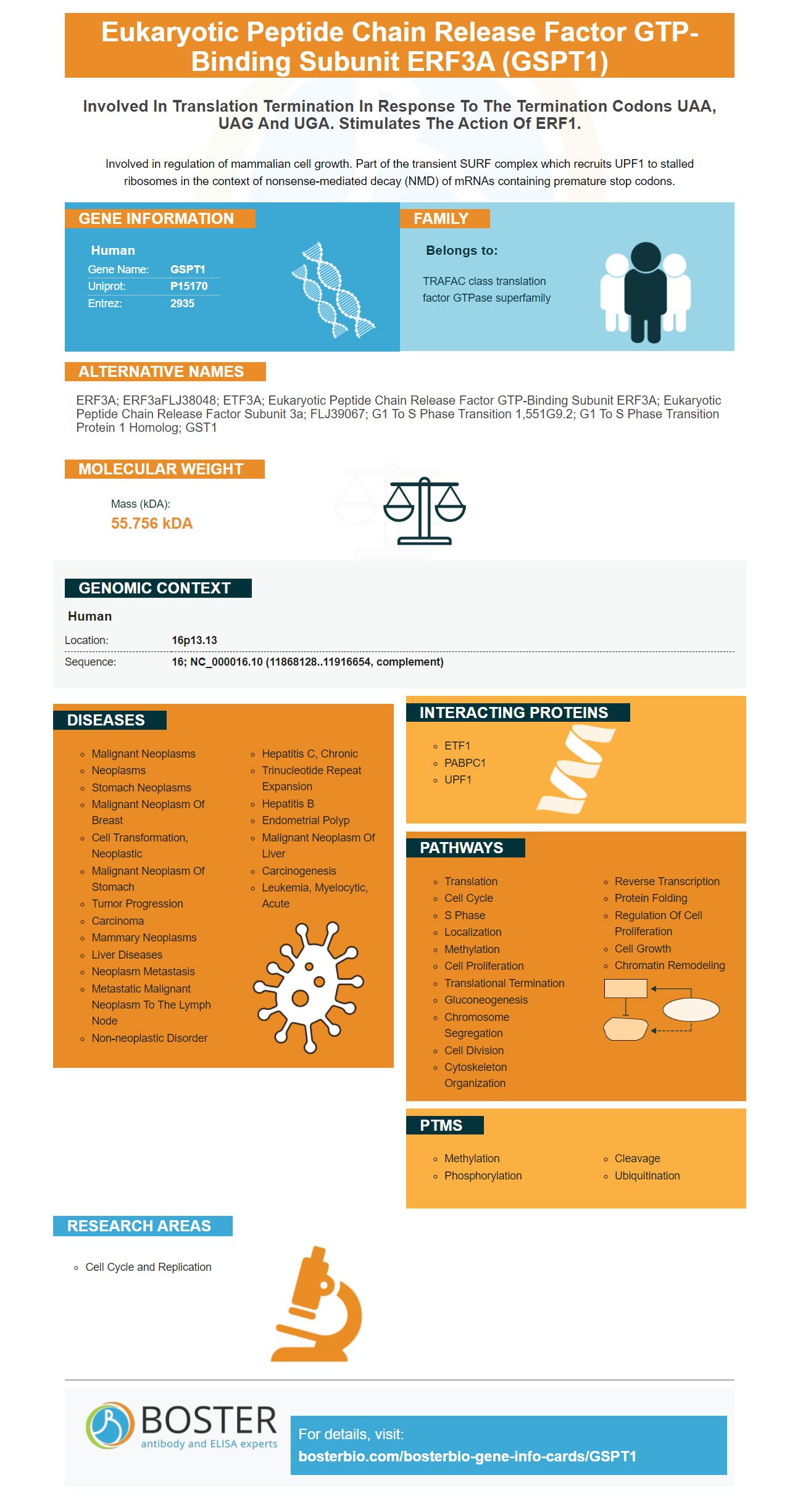
Facts about Eukaryotic peptide chain release factor GTP-binding subunit ERF3A.
Involved in regulation of mammalian cell growth. Part of the transient SURF complex which recruits UPF1 to stalled ribosomes in the context of nonsense-mediated decay (NMD) of mRNAs containing premature stop codons.
Human | |
---|---|
Gene Name: | GSPT1 |
Uniprot: | P15170 |
Entrez: | 2935 |
Belongs to: |
---|
TRAFAC class translation factor GTPase superfamily |
ERF3A; eRF3aFLJ38048; ETF3A; eukaryotic peptide chain release factor GTP-binding subunit ERF3A; Eukaryotic peptide chain release factor subunit 3a; FLJ39067; G1 to S phase transition 1,551G9.2; G1 to S phase transition protein 1 homolog; GST1
Mass (kDA):
55.756 kDA
Human | |
---|---|
Location: | 16p13.13 |
Sequence: | 16; NC_000016.10 (11868128..11916654, complement) |
Anti-GSPT1 antibodies were included in this analysis to study the role of this gene in liver cancer. GO analysis also revealed that anti-GSPT1 antibody significantly decreased pAkt levels and were enriched microglial clusters 2 & 19.
We examined the best uses of GSPT1 as a quantitative, sensitive and specific indicator of gene expression. The Otago Hybridization Buffer contained 0.1% sodium pyrophosphate and 0.5% (w/v.) SDS. It also contains 0.5 mg/ml Heparin. We exposed the Xray film and measured the hybridization signal using a GS-880 Calibrated Densitometer.
Boster Bio's best uses of the GSPT1 mark were evaluated using anti-GSPT1 antibodies as well as other GSPT1 marks. To determine the most efficient antibodies for these markers, three-dimensional gaschromatography was used in conjunction with mass spectrometry.
The BCA method was used to determine the total protein content for each sample. Then, we transferred 1 mg of RNA to PVDF membranes and blocked the blot by adding 5% skimmed Milk. We then exposed the membrane to goat anti-mouse IgG-HRP antibody overnight at 4degC. After blocking, we exposed membranes to goat anti-mouse IgG-HRP antibody overnight at 4°C. We then measured the concentrations of eRF1 and eGFP on an Xray film.
In addition to LOXL1–AS1 downregulation, a miR-708-5p inhibitor (miR-700) completely blocked the inhibitor's effect on cell migration. The inhibitor also increased expression of EGFR, CD44, and other genes that are associated with CRC. This further demonstrates the importance anti-GSPT1 antibodies in the study CRC.
In this study, candidate markers were identified in a subset CSF samples of Parkinson's and Alzheimer disease patients. Normal controls were also included in our research. We found that the protein expression levels of both candidate markers were extremely similar. We can predict the likelihood for both diseases by detecting both markers from separate samples.
For research purposes, GSPT1 is also available commercially. The oral formulation targets both GSPT1 as well as IKZF1/. The company is continuing the study and will soon provide the first dose anti-GSPT1 antibodies in combination chemotherapy. The data will be presented by mid-2018.
We are still not able to fully understand the molecular mechanism that inhibits p-Akt in liver carcinoma. Recent research has revealed that Akt plays a vital role in apoptosis or the inflammation response. This study has suggested that Akt decreases in liver cancer cells and may contribute to its progression. But, further research is needed in order to determine how Akt could be inhibited. This review will examine several possible treatments for liver cancer, and highlight some promising options.
There are two roles in the Akt pathway: to promote cell growth and to promote apoptosis. Akt regulates cell growth by downregulating FoxO transcription factor expression, which in turn induces cyclin-dependent kinase inhibitions (CDK), during the cell cycle process. Hyperactivation results in inhibition in CDKI expression, which leads to cell proliferation. Akt is often highly phosphorylated in the tissues and cells of liver cancer.
Moreover, mice that had Akt knocked out increased their survival rates. The results of this study suggest p-Akt might be a therapeutic target against liver cancer. In a previous study, scientists discovered that liver cancer patients with a pAkt mutation were more likely survive than patients with normal cell levels. The study was published in the journal Oncology.
Akt1 & Akt2 also play roles in liver tumor development. Studies on mice showing that Akt1 and Akt2 proteins are defective in mice have shown an increase in pulmonary metastasis rates. While knocking out p-Akt does not affect the frequency of tumor formation, inhibition of pAkt can increase HCC growth up to 40%.
However, despite the findings from mice, more experiments are needed for further investigation into how these two mechanisms work together in liver cancer. For example, a new drug called aminoquinol inhibits both p-Akt and CDK4/6, which may be used in combination therapy against liver cancer. The study also showed that ADQ can inhibit the activity of MMPs such as MMP-2, MMP-9, and reduce tumor growth in the liver.
ADQ blocks E.cadherin's promoter activity, which is an important factor in cancer development. It also inhibits Twist1 phosphorylation. ADQ also reduces Twist1 expression in different liver cancer cell line types. Further, ADQ inhibits E-cadherin and p-Akt. The drug can also affect cell proliferation, cell motility, apoptosis, and cell motility.
CA also inhibited gene transcription in liver cancer cells in vitro. This drug reduced the amount of apoptotic cell numbers and decreased tumor size in mice. The Huh7R cells were particularly affected by this drug's effect. It also reduced the levels of p–Akt and p–Beclin1, which are hallmarks of apoptosis. Therefore, CA inhibits p-Akt and promotes autophagy in liver cancer.
Genes involved in the process of neurodegeneration are represented by a cluster of genes called microglia. Ex vivo microglia expressed different genes in vitro than ex vivo. This study identified a microglial gene signature, consisting of 1,297 genes, that significantly differed between them. Among these genes are those involved in the innate immune system, pathogen recognition, self-recognition, cell adhesion, and immune signaling.
We performed GO analysis on the LPS-responsive genes in microglia and cells and the corresponding genes in nuclei. The top eight terms were representative to the overall result. Most significantly enriched terms were associated with the inflammatory response of microglia, and there was a high degree of overlap between the cell and the nucleus. It was concluded that GO analysis of microglial clusters 2 and 19 could be useful in studying the pathogenesis of neurodegenerative diseases.
The nuclei and cells of both treatment groups showed high overlap in their clusters. These similarities were reflected within the overall transcriptional modifications. We also assessed whether the data quality affected the results. We also examined whether unique gene count had any effect on the clustering analysis. Both measures had no relationship to clustering. We believe clustering is a better predictor for neurodegenerative disease.
A microglial gene signature linked to multiple diseases is downregulated by chronic EAE mice and upregulated genes related to inflammation, phagocytosis (and lipid metabolism) are upregulated by both models. Additionally, microglia can also express disease-related genes within human brains. Thus, the GO signature of microglia could be indicative of the genetic basis of neurodegenerative disease.
Researchers can identify homeostatic genes in murine and human microglia using a multivariate-RNA-seq method. Sall1, which is associated with gliogenesis, is enriched in human microglia. Microglia's gene signature is made up of more than a thousand genes. GO analysis of these two clusters may reveal other genes that are involved in homeostatic behavior.
A reliable proxy for miRNA expression is the use of frozen CNS tissue to isolate microscopic nuclei. This method may prove useful in the study of neuropathologies. The authors conclude GO analysis 2 and 19 of microglial clusters is a reliable way to identify microglia subpopulations. This type of analysis has many applications, including the study and monitoring of brain inflammation.
PMID: 2511002 by Hoshino S., et al. A human homologue of the yeast GST1 gene codes for a GTP-binding protein and is expressed in a proliferation-dependent manner in mammalian cells.
PMID: 19417104 by Yamashita A., et al. SMG-8 and SMG-9, two novel subunits of the SMG-1 complex, regulate remodeling of the mRNA surveillance complex during nonsense-mediated mRNA decay.