This website uses cookies to ensure you get the best experience on our website.
- Table of Contents
1 Citations 3 Q&As
2 Citations
Facts about Gamma-aminobutyric acid receptor-associated protein.
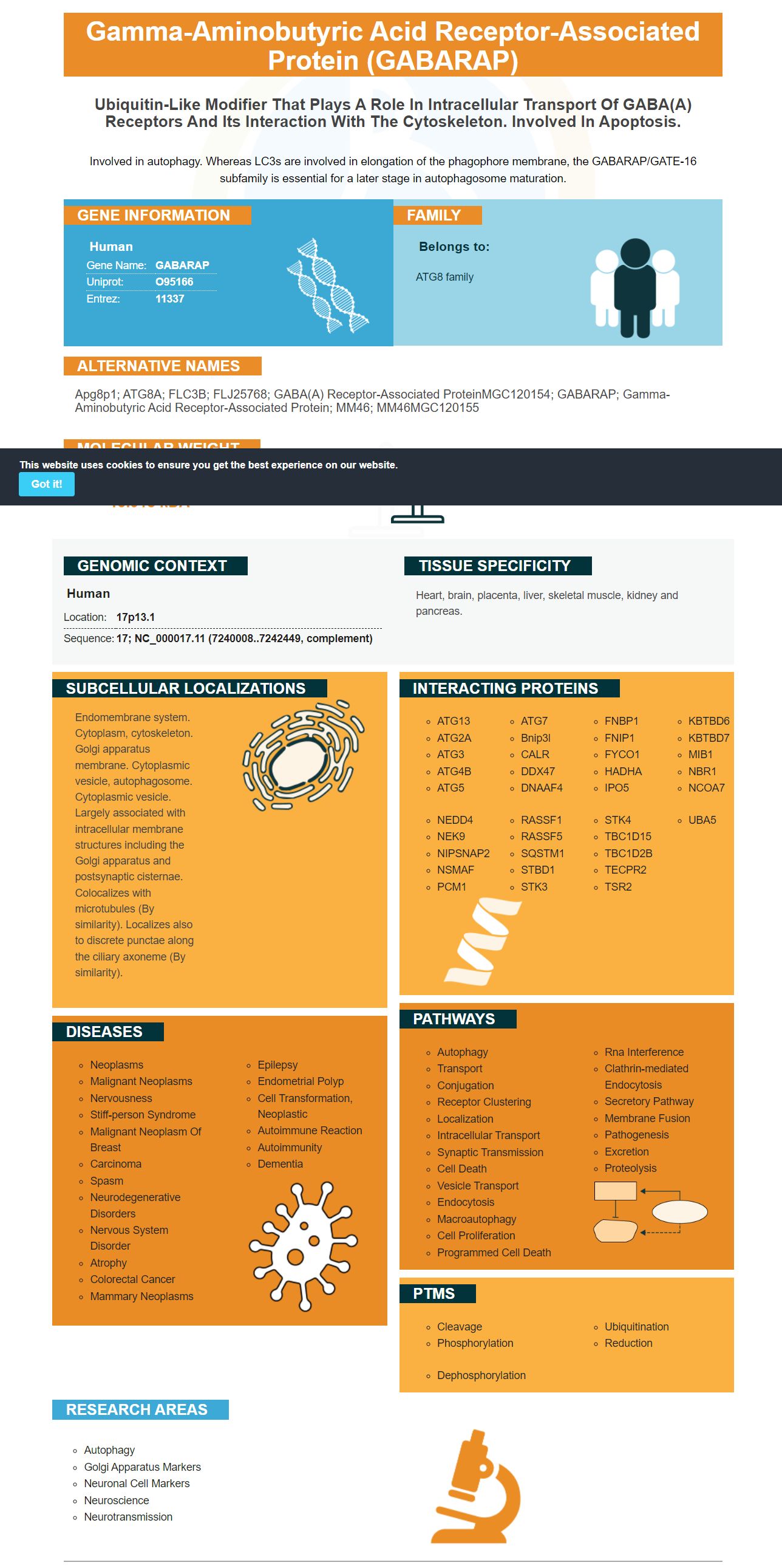
Involved in autophagy. Whereas LC3s are involved in elongation of the phagophore membrane, the GABARAP/GATE-16 subfamily is essential for a later stage in autophagosome maturation.
Human | |
---|---|
Gene Name: | GABARAP |
Uniprot: | O95166 |
Entrez: | 11337 |
Belongs to: |
---|
ATG8 family |
Apg8p1; ATG8A; FLC3B; FLJ25768; GABA(A) receptor-associated proteinMGC120154; GABARAP; gamma-aminobutyric acid receptor-associated protein; MM46; MM46MGC120155
Mass (kDA):
13.918 kDA
Human | |
---|---|
Location: | 17p13.1 |
Sequence: | 17; NC_000017.11 (7240008..7242449, complement) |
Heart, brain, placenta, liver, skeletal muscle, kidney and pancreas.
Endomembrane system. Cytoplasm, cytoskeleton. Golgi apparatus membrane. Cytoplasmic vesicle, autophagosome. Cytoplasmic vesicle. Largely associated with intracellular membrane structures including the Golgi apparatus and postsynaptic cisternae. Colocalizes with microtubules (By similarity). Localizes also to discrete punctae along the ciliary axoneme (By similarity).
The anti-GABARAP antibody (8H5) can discriminate GABARAP from its relatives, making it suitable for IF-labelling of tissue sections. Here are some uses for the anti-GABARAP antibody. They can also be applied to species-specific applications. In this article, we will discuss the advantages and disadvantages of using this antibody. Also, we will discuss its image analysis.
The anti-GABARAP (8H5) antibody was used in immunostaining. The antibody demonstrated good selectivity and a high signal-to-noise ratio. It recognized residues spanning positions 7 to 18 and a high affinity toward the antigen. These positive characteristics indicate that this antibody is a promising tool for immunostaining. Its high affinity may be useful for the diagnosis and detection of various neurological diseases.
GABARAP is a member of the adaptor-like protein family. These proteins share similar but partly overlapping features. First described as a marker for type-A GABA receptors in neurons, the protein is now implicated in several intracellular transport processes. In addition to its role in fusion, the GABARAP marker also plays a pivotal role in autophagy, an evolutionarily conserved process. It can undergo lipidation, which facilitates its association with autophagosomal membranes. The GABARAP marker likely also associates with tubule vesicular structures.
As with other ATG8 members, elucidating their unique roles is challenging. Fluorescent protein (FP)-tagged ATG8 reporters have provided multiple new insights. However, this technique may also be accompanied by steric hindrance and artifacts. Additionally, the available antibodies against diverse ATG8 members often show cross-reactivity and are not transparent to the user.
This anti-GABARAP (8H5) antibody was designed to detect endogenous GABARAP within some, but not all, LC3B+ structures. The GABARAP epitope overlaps with a-helices 1 and 2 and extends across the ubiquitin-like fold. The antibody binds to these non-conserved residues, defining the binding specificity of this antibody to GABARAP.
In recent years, scientists have attempted to analyze the specificity of the GABARAP antibody by using plasmids encoding YFP-GABARAPL1 or CFP-GABARAPL2 to detect this protein. The results showed that the transfected cells exhibited fluorescence exclusively in channels corresponding to cyan fluorescent protein, YFP, or CFP.
ATG4 and ATG3 process GABARAP's precursor form. In addition, GABARAP is found in the brain and heart, where it is involved in autophagy. Anti-GABARAP (8H5) antibody has been used successfully in IF-labelling of tissue sections. It can also be used for immunostaining to detect protein concentrations in different tissues.
The use of this antibody requires highly specific antibodies. Using multiplex chromatography, researchers may increase protein detection frequency by up to 20%. The chromatography technique also involves biotinylation and immunoprecipitation. For cellular proteins, each type of antibody is attached to a different colored bead. This enables researchers to perform immunofluorescence staining of tissue sections.
IF-labelling of tissue sections can be used to visualize the association of GABAA receptors with autophagosomes. The protein is a highly conserved member of the ATG8 family. It has been implicated in numerous intracellular transport processes and is essential in autophagy, an evolutionarily conserved process. The GABARAP is lipidated, promoting its association with autophagosomal membranes.
GABARAP is also secreted in EVs. It has been suggested that the EVs contain RBPs and mitochondrial contents. The APEX2-mediated proximity labelling method may be useful in the discovery of other proteins in EVs. However, the mechanism of secretion is unknown. IF-labelling of tissue sections using this antibody should be further studied in detail.
APEX2-GABARAP was also tested in EVs. EV lysates were incubated with streptavidin-coated beads and washed. The eluate was then collected, where biotinylated EV proteins were detected. These are the two methods used in IF-labelling of tissue sections.
The primary anti-GABARAP (8H5) antibody is a monoclonal antibody that produces intracellular staining in a variety of cell types. The secondary antibody was diluted in TBS-T and added to the wells. The secondary antibody was then added and incubated for a further 60 min. Afterward, the immunoreactive cells were visualized and stained with the primary antibody.
Using this monoclonal anti-GABARAP antibody can be useful in IF-labelling of tissue slices. GABARAP is secreted in extracellular vesicles and can be detected using immunoblotting. Additionally, a cellular impurity marker, CD81, was used in the EV sample to identify EVs.
Using anti-GABARAP (8H5) as the immunogen, we examined the staining patterns of GABARAP in human glioblastoma cell lines. The anti-GABARAP antibody detects GABARAP in the cytoplasmic vesicles of cells stained with NorthernLights(tm) 557-conjugated anti-Rat IgG secondary antibody and DAPI.
GABARAP is a member of the adaptor-like protein (ATG) family with a wide action spectrum and partially overlapping features. Originally identified as a receptor tyrosine kinase in neurons, GABARAP is now implicated in numerous intracellular transport processes, including autophagy, an evolutionary conserved process. GABARAP also undergoes lipidation, promoting association with autophagosomal membranes and tubule vesicles.
In addition to recognizing GABARAP, the antibody was able to detect lipidated YFP-GABARAP, a protein that is associated with autophagosomes. The lipidated GABARAP protein accumulates in the cytoplasm, and confocal imaging applications reveal bright puncta. In HAP1 KO cells, however, the antibody detected a faint background stain. Nevertheless, this result indicates that anti-GABARAP antibody is highly specific.
The binding specificity of the anti-GABARAP (8H5) antibodies was confirmed by array-based oligo-peptide scanning. The anti-GABARAP antibody recognized residues from position seven to 18 in both IF and western blotting, with the fourth mer showing the strongest signal. Thus, anti-GABARAP (8H5) antibodies are more specific against GABARAP, which makes them the most appropriate choice for immunostaining.
The antibodies were used in an ELISA to analyze the immunostaining of a cell line. In this assay, mouse anti-GABARAP (8H5) antibody was used as the capture and detection antibodies. Then, a monoclonal anti-rat IgG antibody was used as a negative control. Finally, supernatant from the hybridoma cells was diluted 1:10. Following the wash steps, the secondary antibodies were added and visualized by horseradish peroxidase-coupled mouse monoclonal anti-rat IgG heavy chain. The absorbance of the secondary antibodies was measured at 650 nm.
The specificity of an antibody is highly dependent on the type of immunogen. Synthetic peptides or purified proteins are used as immunogens. Moreover, antibody specificity is also dependent on the structure of the immunogen. This is the reason why it is crucial to use appropriate dilutions and protocols when using an antibody. The datasheet should describe all relevant information and precautions.
PMID: 9892355 by Wang H., et al. GABA(A)-receptor-associated protein links GABA(A) receptors and the cytoskeleton.
PMID: 11146101 by Okazaki N., et al. Interaction of the Unc-51-like kinase and microtubule-associated protein light chain 3 related proteins in the brain: possible role of vesicular transport in axonal elongation.
*More publications can be found for each product on its corresponding product page