This website uses cookies to ensure you get the best experience on our website.
- Table of Contents
Facts about Phospholemman.
Associates with and Modulates the activity of This sodium/potassium-transporting ATPase (NKA) which transports Na(+) from the cell and K(+) into the cell.
Inhibits NKA activity in its unphosphorylated state and stimulates activity when phosphorylated.Reduce glutathionylation of the NKA beta-1 subunit ATP1B1, thus reversing glutathionylation-mediated inhibition of ATP1B1. Contributes to female sexual development by maintaining the excitability of neurons that secrete gonadotropin-releasing hormone.
Human | |
---|---|
Gene Name: | FXYD1 |
Uniprot: | O00168 |
Entrez: | 5348 |
Belongs to: |
---|
FXYD family |
FXYD domain containing ion transport regulator 1; FXYD domain-containing ion transport regulator 1; MGC44983; PLMphospholemman
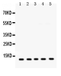
Mass (kDA):
10.441 kDA
Human | |
---|---|
Location: | 19q13.12 |
Sequence: | 19; NC_000019.10 (35137206..35143109) |
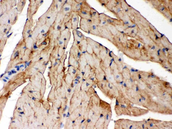
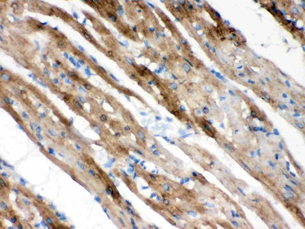
Highest expression in skeletal muscle and heart. Moderate levels in brain, placenta, lung, liver, pancreas, uterus, bladder, prostate, small intestine and colon with mucosal lining. Very low levels in kidney, colon and small intestine without mucosa, prostate without endothelial lining, spleen, and testis.
Cell membrane, sarcolemma; Single-pass type I membrane protein. Apical cell membrane; Single-pass type I membrane protein. Membrane, caveola. Cell membrane, sarcolemma, T-tubule. Detected in the apical cell membrane in brain. In myocytes, localizes to sarcolemma, t-tubules and intercalated disks.
If you're looking for information on Steven Boster, you've come to the right place. This article discusses his research, publications, and career. You'll learn about Boster Bio's high affinity primary antibodies for the FXYD1 marker. These antibodies are cited widely and have been in use for over 25 years. They've been used in a variety of research applications.
Researchers have developed a new evolutionary scenario for the FXYD1 gene and have identified the genes and protein sequences for seven different FXYD molecules. These genes and proteins were first identified in rodents and humans in 2000. These proteins are found predominantly in the proximal tubules, medullary thick ascending limbs, and CCD principal cells. The gene FXYD1 also colocalizes with Na,K-ATPase.
The MeCP2 gene codes for a transcriptional regulator. Overexpression of this gene is associated with the pathogenesis of Rett syndrome (RTT), a neurodevelopmental disorder. It is unknown if the FXYD1 subunit is affected by IGF-1, but the gene is overexpressed in RTT. The FXYD1 marker has also been implicated in the pathogenesis of RTT.
The gene encodes a 72 amino acid protein that belongs to the FXYD family of ion transport regulators. The FXYD motif is known to have evolved in the earliest vertebrates, and the first known example of an FXYD protein is present in the sea lamprey. There have been no non-vertebrate homologs found. Researchers have thus been able to analyze the role of this protein in many cellular processes, including the development of the brain.
A recent study has found that IGF-1 treatment reduced FXYD1 protein expression significantly in RTT mice. IGF-1 treatment also reduced the expression of mTOR and S6 in these RTT mice. These results indicate that FXYD1 is a key component of the brain, and the treatment of this gene with the IGF-1 hormone may have therapeutic implications for brain disorders. This gene is highly expressed in the rat, but it is still unknown how it affects cognitive and motor function in humans.
The protein phosphorylation of FXYD1 inhibits the activity of the PI3K/AKT signalling pathway, and the effects of IGF-1 treatment were observed in RTT mice. It is important to note, however, that the treatment of IGF-1 also increased p-FXYD1 levels. In addition, the effects of IGF-1 treatment on FXYD1 and p-mTOR in the mice have been found to be reversible.
Recently, a new study has described a gene called FXYD1 that is involved in the protection of eNOS from oxidative stress and dysregulated redox signaling. The gene is intimately related to eNOS and the production of superoxide. The research has potential therapeutic implications in both hypertension and diabetes. Further studies of FXYD1 and its metabolites should focus on identifying novel therapeutic targets.
The gene product FXYD1 is expressed in various tissues throughout the body. It plays a central role in the regulation of Na,K-ATPase activity and is expressed in various types of tissues including the brain, uterus, liver, and placenta. The gene also induces Cl-selective currents and has been identified as a tissue-specific regulator of Na,K-ATPase. The protein inhibits the activity of NKA when unphosphorylated and stimulates activity when phosphorylated.
The FXYD1 protein has a highly organized secondary structure similar to that of genes. Intron-exon junctions are the locations where the proteins form the structure of the gene. The secondary structure of FXYD1 is summarized in Figure 2B. A transmembrane helix (H2) precedes a short helical segment (h2). The short helix (H3) spans the basic RRCRCK sequence.
The structure of FXYD1 is shown in a membrane frame. The structure is oriented at a 15deg angle to satisfy the transmembrane tilt. Interestingly, the helical helix is nearly parallel to the membrane axis. It is thought that the helix reorientation from the membrane surface is triggered by phosphorylation, which introduces repulsive negative charges at S63 and S68 of H4.
In addition to the phenotypic data, a new study has provided further insight into the mechanism by which FXYD1 binds to the Na,K ATPase subunit. The results indicate that a phosphorylated version of FXYD1 interacts with the subunit's cytoplasmic domain. A study of FXYD1 has also provided insight into the association of FXYD1 with subunits of the enzyme, and it has paved the way for future structural analysis of enzyme complexes.
The phosphorylated form of FXYD1 contains a phenylalanine residue called Phe28 that protrudes out of the transmembrane helix. The residues Gly20, Gly31, and Gly25 are fully conserved and represent the three Ser residues and one Thr69, respectively. The surface color code reflects the electrostatic potential of the residues. Red is the -8kBT whereas blue is the +8kBT. The FXYD1 structure is viewed 90 degrees around the membrane's Y axis. The transmembrane helix demonstrates predicted interactions with Na,K-ATPase a subunit.
The FXYD1 gene is expressed in electrically excitable tissues that specialize in transport. The six mammalian members of the FXYD family interact with Na,K-ATPase, and their shark homolog modulates its rate constant and affinity for Na. A study in rats also suggests that FXYD1 binds to Na,K-ATPase-A, a subunit of the Na,K-ATPase.
The Mn PRE profile is a useful tool for understanding the dynamics of FXYD1 proteins. This model uses the structure of FXYD1 molecules to determine their relative positions in membranes. It also provides an insight into the structure of FXYD1 in micelles. It has been shown that the Mn PRE profile has a remarkably similar profile to that of h2-H3, with the exception of the length of the helix H4.
PMID: 9169143 by Chen L.-S.K., et al. Characterization of the human and rat phospholemman (PLM) cDNAs and localization of the human PLM gene to chromosome 19q13.1.
PMID: 10950925 by Sweadner K.J., et al. The FXYD gene family of small ion transport regulators or channels: cDNA sequence, protein signature sequence, and expression.