This website uses cookies to ensure you get the best experience on our website.
- Table of Contents
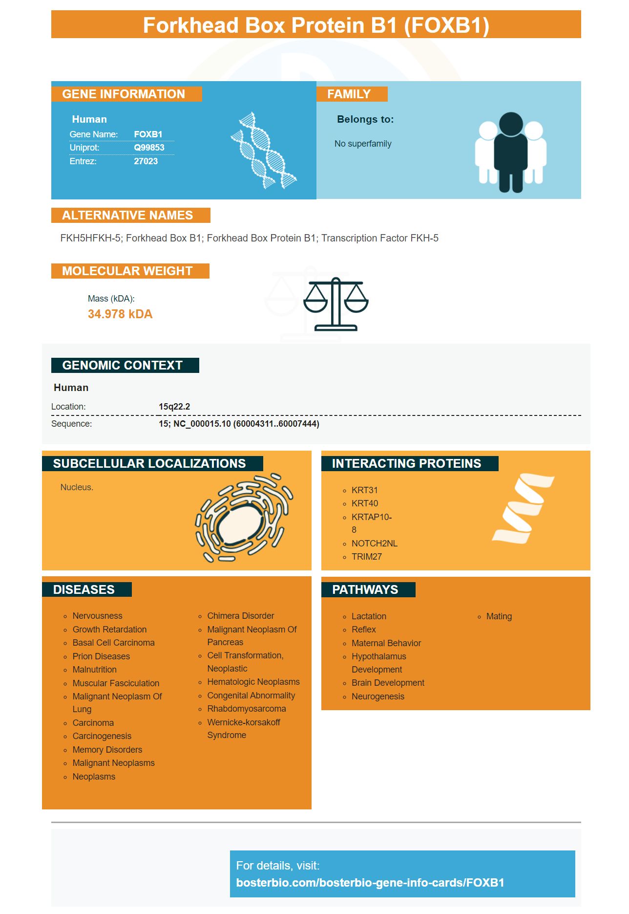
Facts about Forkhead box protein B1.
Human | |
---|---|
Gene Name: | FOXB1 |
Uniprot: | Q99853 |
Entrez: | 27023 |
Belongs to: |
---|
No superfamily |
FKH5HFKH-5; forkhead box B1; forkhead box protein B1; Transcription factor FKH-5
Mass (kDA):
34.978 kDA
Human | |
---|---|
Location: | 15q22.2 |
Sequence: | 15; NC_000015.10 (60004311..60007444) |
Nucleus.
This article will provide an overview of FOXB1 marker information. It also discusses B1 of the Forkhead protein, KHDRBS2 (and TRA-1-60 (+), as well as the role of KHDRBS2 cells and TRA-1-60 (+). You can also read the rest to learn more about these markers.
Boster Bio can provide a very accurate diagnosis of cancer by using Forkhead box gene expression from Boster Bio. Forkheadbox gene expression is a biomarker of mucosa associated lymphoid tissue Lymphomas, which can have poor prognosis. Expression of Forkhead box gene expression predicts transformation to diffuse large B-cell lymphoma.
This test uses microRNA-376a, which targets Forkhead protein P2. Liu R is responsible for controlling NF-kappaB positive feedback loops. It also inhibits osteosarcoma cellular growth. Regulating p53-dependent DNA damaging responses is crucially important for Forkhead Box Protein B1. Baltimore D also suppresses tumorigenesis through inhibiting FOXP3 and B lymphocyte development.
Foxp3 is a transcription factor that controls the expression of Forkhead Box genes. This transcription factor also regulates CIP1 as well as p21WAF1. This means that Forkhead transcript factor can be a key regulator of these proteins. The key component of osteosarcoma-cell growth arrest is Forkheadbox gene expression. This marker can be used for screening for osteosarcoma or its associated pathologies.
The expression of FOXh2 in human iPSCs will determine the best use of this marker. It is necessary to define the anterior primitive streak in early development. It is also required for the maturation and activation if late pluripotency marker activation, as well as the transition from mesenchymal into epithelial states. This is important because somatic cells in human bodies experience a temporary state similar to mesendoderm.
The best use of FOXh2 is in reprogramming iPSCs. This gene promotes downregulation CD13 and promotes EpCAM, ZFP42 and endogenous SOX2. The gene also has the ability to facilitate the maturation of TRA-1-60 (+) cells. FOXh2 may also promote differentiation of T-cells derived from splenic tissue.
FOXB1 is necessary for induction of OL in zebrafish. It regulates OL differentiation and generation. It also inhibits OPC proliferation. These functions make FOXB1 a valuable marker in stem cell research and cell biology. But how do you make the most of the FOXB1 gene? We examine the roles of the FOXB1 genes in neuronal development.
Cortical VZ contains the FOXB1 protein, which is functionally homogeneous. It is not clear what role this gene plays in OL development. However, it is worth pointing out that KHDRBS2 is a novel protein in the OL. FOXB1 is also expressed in neuroepithelial tissues such as ovaries, uterus, and uterus.
It can also be used to study ischemic stroke. The FOXB1 gene is abundantly expressed in the Thalamus, an area of the forebrain containing high Foxb1 levels. KHDRBS2 expression in Astrocytes is also important for understanding FOXB1's role in the ischemic attack.
We used TRA-1-60 (+ cells as the source of gene expression analysis in this study. We isolated TRA-1-60 (+), cells over a period of time and compared their gene expression profiles with iPSCs. Our analysis showed that there were transient changes of GDF3, LEFTY2, NODAL and GDF3 in the TRA-1-60 (+). cells, as well as in iPSCs.
TRA-1-60 (+), cells were found in our study on day 7. Their ability to form iPSC colonies increased and they were detected as early as day 7. TRA-1-60 (+ cell) showed a reprogramming performance comparable to ESCs, iPSCs, and on day 20, TRA-1-60 (+ cell) had a similar level of efficiency. Furthermore, this study has shown that TRA-1-60 (+) cells can differentiate into various cell types and tissues.
We used TRA-1-60 to determine if human embryonic cells stem cells were pluripotent. GloLIVE NL493 conjugated human anti-mouse TRA-1-60 monoclonal antibodies was used to visualize TRA-1-60 in live BG01V embryonic stem cells. Hoechst 33442 was used as a counterstain to the nuclei. Positive staining of TRA-1-60 (+) cells in human stem cell cultures indicates pluripotency.
In addition to tracheal cancer, tracheal tumors, and ovaries, the FOXB1 marker is involved in brain development. However, it is still not clear if the marker plays any role in OL-development. It is possible to use TRA-1-60 (+ cells as a source of OL replacement therapy. The FOXB1 marker works only when the FOXB1 gene is expressed in cells.
The FOXB1 marker is used to detect the lineage for neurons and astrocytes, as well as cancer cell lines. They have high levels myelin fundamental protein and hPLAP. Claudin11 was also colocalized to hPLAP as well as g-galactosidase. Foxb1 expression was found to be colocalized in mature oligodendrocytes with characteristic lamellae and morphology.
The critical role of the FOXB1 mark in differentiation of iPSCs has been identified as a crucial component of standardization and validation for iPSC technology. The marker has been identified to be a crucial component in the differentiation human ESCs. This study also showed that iPSCs can produce cardiomyocytes. Moreover, the expression of several cardiomyocyte-associated genes was downregulated after differentiation. Moreover teratomas could also be formed by iPSCs derived from immunodeficient mice. Teratomas represent a landmark in pluripotency research as they are tumors that contain tissue of three germ layers.
Researchers removed certain factors one by one from the pool of 24 cells to achieve ESC-like colonies. They eventually identified four key factors: Oct4, Sox2, Klf4, cMyc. The transcriptional profiles of the downregulated group were similar to those of the ESC-like colonies that were obtained via retroviral transfer. Consequently, iPS-derived stem cells have a high proportion of induced gene.
Gdf3 represents another gene that is expressed in human embryonic-stem cells. It is essential for the formation of gastrula mesoderm, and endoderm. It is also a cofactor for the Nodal Signaling System in Zebrafish and Crucian Carp embryos. Gdf3 is a useful marker to differentiate iPSCs.
Reprogramming adult cells for iPSCs is a risky business. The viruses used for manipulating cells may trigger the expression oncogenes. Scientists have recently revealed a new method to eliminate oncogenes by inducing pluripotency. This discovery will improve the use of iPS cells in treating human diseases. Researchers can now avoid the potential risks associated to c-Myc by using this new technique. It also increased chances of creating chimeras having cancer-resistant properties.
PMID: 14702039 by Ota T., et al. Complete sequencing and characterization of 21,243 full-length human cDNAs.