This website uses cookies to ensure you get the best experience on our website.
- Table of Contents
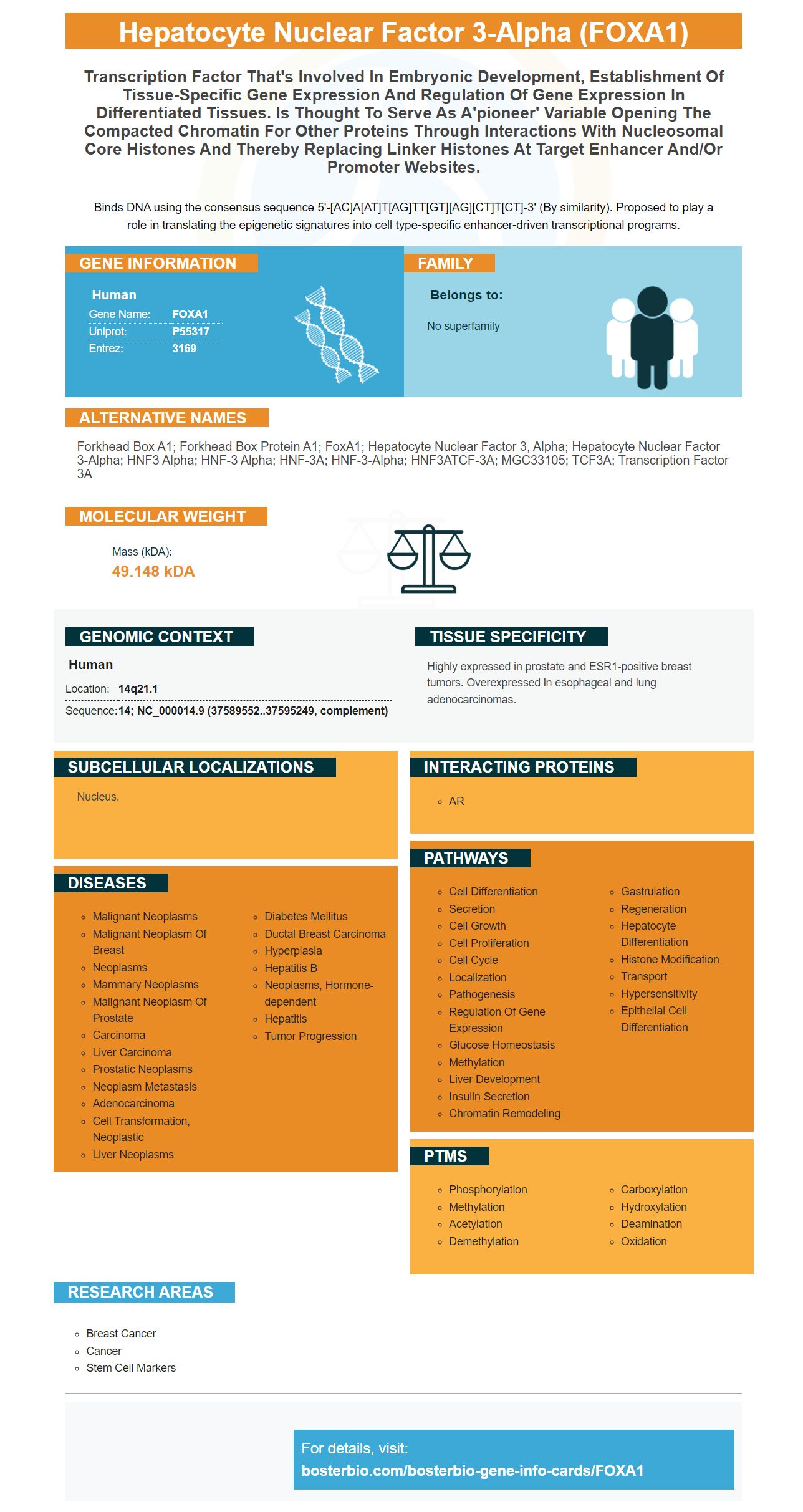
Facts about Hepatocyte nuclear factor 3-alpha.
Binds DNA using the consensus sequence 5'-[AC]A[AT]T[AG]TT[GT][AG][CT]T[CT]-3' (By similarity). Proposed to play a role in translating the epigenetic signatures into cell type-specific enhancer-driven transcriptional programs.
Human | |
---|---|
Gene Name: | FOXA1 |
Uniprot: | P55317 |
Entrez: | 3169 |
Belongs to: |
---|
No superfamily |
forkhead box A1; Forkhead box protein A1; FoxA1; hepatocyte nuclear factor 3, alpha; hepatocyte nuclear factor 3-alpha; HNF3 alpha; HNF-3 alpha; HNF-3A; HNF-3-alpha; HNF3ATCF-3A; MGC33105; TCF3A; Transcription factor 3A
Mass (kDA):
49.148 kDA
Human | |
---|---|
Location: | 14q21.1 |
Sequence: | 14; NC_000014.9 (37589552..37595249, complement) |
Highly expressed in prostate and ESR1-positive breast tumors. Overexpressed in esophageal and lung adenocarcinomas.
Nucleus.
The FOXA1 gene is a significant pioneer among transcription factors. This gene may prove useful in stratifying patients with ER+ breast cancer or as a second-line treatment for prostate cancers that are resistant to drugs. Further research into the role of FoxA1 in the clinic is needed to comprehend its potential clinical advantages. Here are some of its most valuable uses:
The FoxA1 transcription factor is a pioneering element in the transcriptional process. It engages with DNA's condensedchromatin to aid in the association of other transcription factors. Its "winged hexagon" structure allows it access to the major groove of DNA. Its alpha-helix is made up of two loops, one on each side. The wings also create an additional site-specific contact. Its structure is similar to that of Histone h2.
It isn't known what role FOXA1 plays in ER-negative disorders. However, it is associated with the growth ER positive and ER–AR+ prostate cancer cell lines. Thus targeting this gene could be an appealing second-line treatment option for patients suffering from cancer who are not candidates for conventional chemotherapy or radiation therapy. It is also present in only a few adult tissues, making it an ideal second-line treatment.
FOXA1 is a key player in the development of hormone-dependent cancers. This is particularly true for prostate and breast cancers. Its function is to determine where and how transcription factors are able to bind to DNA and this protein is vital to the function of nuclear receptors. Numerous studies have demonstrated that this transcription factor can be used as a biomarker for identifying all types of cancer. It may also be a sign that there is abnormal cell growth as it is present in both prostate and breast cancers.
A recent study suggests that FoxA1 is closely linked to the AR gene, which has significant roles in hormone-resistant prostate cancers. Furthermore, metastases from prostate and breast cancers have been identified to contain more FoxA1, indicating the existence of a shared dependency on this transcription factor at secondary sites. This could also indicate an alternative mechanism to activate the AR in CRPC.
The study has a number of implications for the use of the FOXA1 marker in breast cancer research. It was discovered that FOXA1 binding is affected by more than half the risk-associated SNPs. As such, it is important to study other transcription factors to determine their significance in the risk of breast cancer. The study also demonstrates an association between FOXA1 mutation and the p53-mutation.
In the selection of treatment options, it may be beneficial to consider the ER status in breast cancer as a prognostic indicator. In one study, ER positivity was associated with poorer outcomes for patients suffering from ER+ breast cancer and LN-negative cancer. This is especially relevant for larger tumors. This marker can also help doctors predict the progression of the disease and determine the best follow up strategy.
Surprisingly, nearly all breast cancers express ER. Furthermore, the majority of them have the ER+ phenotype, which is tolerant to ER inhibition. Previously, Ramos et al. Ramos and al. earlier reported that hypermethylation (or overmethylation) of ESR1 could be a poor indicator of prognosis in the case of sporadic breast cancer. However, more studies are required to determine if the expression of ESR1 is really a reliable indicator of prognosis or not.
The survival rate for metastatic disease has been linked to Ki-67 expression. Ki-67 expression is highly increased in hyperplastic areas and adjacent normal terminal duct lobular units. High risk of developing breast carcinoma is also linked to Ki-67. Ki-67 is associated to ER expression which is affected in the carcinogenesis process. Patients with high Ki67 activity in early stage breast cancer have a greater chance of recurrence and a poorer survival rate. The International Ki-67 Breast Cancer Working Group recognizes the marker's limited clinical validity.
BCL2 protein expression could be a powerful prognostic indicator for breast cancer. The prospective study of more than 11 000 women with early stage breast cancer suggests that BCL2 expression could be a reliable and reliable prognostic marker in ER+ patients. Although the clinical value isn't evident, this study confirms the significance of BCL2 expression in breast cancer survivorship.
However, despite the absence of clinical value, the ER+ survival rate is an unproven indicator. While the MFS in ER+ disease is similar to that of N-types but the mortality rate of N-type patients is significantly higher. However, BCL2 protein expression may not provide the same prognostic data. This is why ER positivity a poor indicator of prognosis for ER+ breast carcinoma.
A new study suggests that a particular gene, Boster Bio could be a biomarker for ER+ HER2 disorder. The biomarker was discovered by the use of RNA sequencing. To determine if it was able to determine the effectiveness of treatment the researchers looked at breast cancer patients' blood samples. Patients with breast cancer were divided by the HER2 level. The researchers found that patients in the T-DM1 arm had a higher chance of achieving a pCR than those in the trastuzumab-and-paclitaxel-group.
The study also showed that triple-agent combinations had higher pCR rates as compared to doublet arms T+ D and P-D. This suggests that patients suffering from HER2-positive cancer may not require chemotherapy during neoadjuvant therapies. In addition, this study suggested that simple double-targeted therapy may produce satisfactory results. In addition, the results suggest that the drug bosterazumab does not have any effect on heart toxicity, despite the fact that a decrease in dose dependent EF was reported.
Furthermore, TP53 and MYC gene family interactions are linked to rapid progression of disease. A whole-genome sequence study also found a significant increase in somatic mutational burden post-therapy. TP53 and DYNC1h2 mutations were often observed in SHH-MBs. In addition, patients who did not respond to the first chemotherapy regimen showed higher rates of TP53 and MYCN amplifying.
In addition to Boster Bio, researchers have also identified an additional gene, MYC messenger RNA. This gene is found in more than 50 different tumor types, including ER+ HER2 as well as MYC-amplified tumors. Its identification in children suffering from neuroectodermal tumors can help doctors differentiate between different kinds of disease.
In addition to its potential as a biomarker T-DM1 sensitivity is also correlated with the expression of HER2, although the relationship was weaker than with T-DM1 sensitivity. T-DM1 sensitivity was also linked to RAB5A and HER2 as biomarkers for TDM1-mediated cell responses.
One study showed that prostate cancer patients with high FOXA1 levels had lower rates of survival without relapse. However, it did not provide an independent prognostic indicator when utilized in a Cox regression analysis in conjunction with preoperative PSA level, pT stage, Gleason score, and margin status. These results suggest that FOXA1 expression may not be as helpful as previously thought.
Numerous studies have demonstrated that FOXA1 expression is related to the amount of epithelial AR. They also discovered that the two markers had excellent concordance in the immunoreactivity of human prostatic tissues. Further research is required to determine whether FOXA1 can be utilized as a second-line treatment for prostate cancers that are resistant to treatment. The study also suggests that FOXA1 could be used as an additional marker in the detection of drug-resistant prostate cancer.
The mRNA level of FOXA1 was determined by QPCR. Western Blot was used to determine the protein level. Both types of cells were cultured in T75-cell-culture flasks until confluence was 70-80%. In addition, the cell line was transfected either nonspecific siRNA or one of three different types of siRNAs. The cells were then incubated for 72 hours.
An alternative is to examine the tumors of patients who have FOXA1 and to identify them. This is crucial as FOXA1 is associated with the p27 gene in primary prostate cancer. FOXA1 knockdown inhibits PC-3 cell growth. It also induces G1 stoppage in PC-3 cells. This marker can be used to aid in the development of an alternative treatment for drug-resistant prostate carcinoma.
The expression of FOXA1 is closely associated with the AR and the steroid hormone receptors. The expression of FOXA1 increased when tumors grew and metastasized which suggests a role in the hormone-resistant prostate cancer. Patients who have high FOXA1 expression could be more responsive to hormone therapy. This new marker may provide a novel way to treat patients suffering from CRPC.
It is not clear if FOXA1 is a viable second-line treatment for drug-resistant prostate carcinoma. However, the results suggest that the protein is relevant in prostate cancers that are drug-resistant. This marker should be further investigated as an alternative to treatment for prostate cancers that are resistant to treatment. It should be possible to detect patients with FOXA1 in their tumors with a simple blood tests.
PMID: 8652662 by Bingle C.D., et al. Molecular cloning of the forkhead transcription factor HNF-3 alpha from a human pulmonary adenocarcinoma cell line.
PMID: 10899756 by Navas M.A., et al. The human HNF-3 genes: cloning, partial sequence and mutation screening in patients with impaired glucose homeostasis.