This website uses cookies to ensure you get the best experience on our website.
- Table of Contents
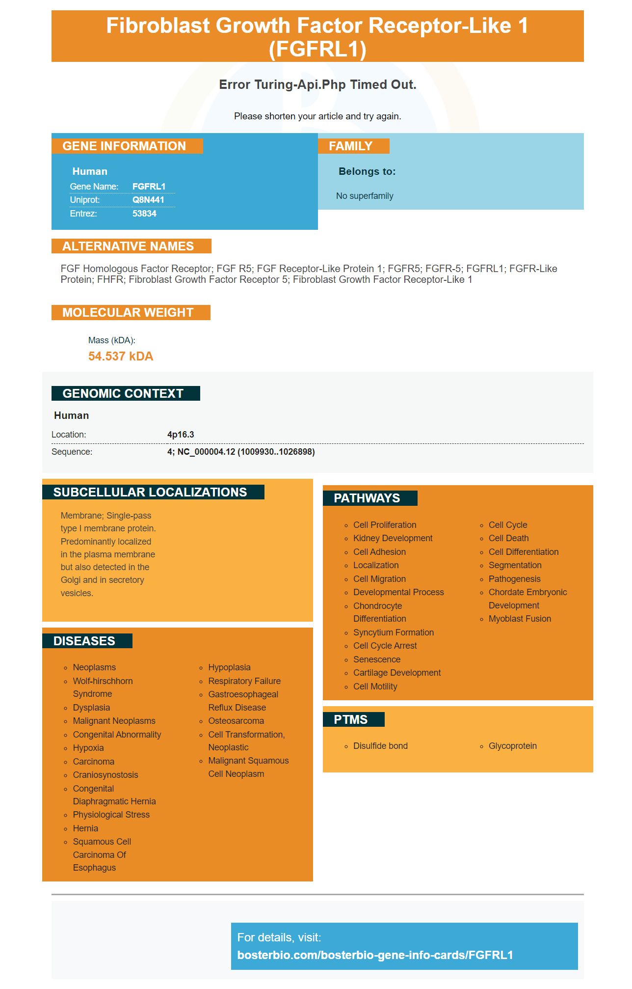
Facts about Fibroblast growth factor receptor-like 1.
Please shorten your article and try again.
Human | |
---|---|
Gene Name: | FGFRL1 |
Uniprot: | Q8N441 |
Entrez: | 53834 |
Belongs to: |
---|
No superfamily |
FGF homologous factor receptor; FGF R5; FGF receptor-like protein 1; FGFR5; FGFR-5; FGFRL1; FGFR-like protein; FHFR; Fibroblast growth factor receptor 5; fibroblast growth factor receptor-like 1
Mass (kDA):
54.537 kDA
Human | |
---|---|
Location: | 4p16.3 |
Sequence: | 4; NC_000004.12 (1009930..1026898) |
Membrane; Single-pass type I membrane protein. Predominantly localized in the plasma membrane but also detected in the Golgi and in secretory vesicles.
The FGFRL1 biomarker for growth factor receptor-like 1 is a protein with many biological functions. This protein is involved with the regulation of gene expression in a study on stem cells. Boster Bio provides researchers with the FGFRL1 antibody and the Cy3-conjugated goat anti-goat IgG secondary antibody. In a related study, the FGFRL1 marker was used to identify cancer stem cells in mice.
This boster bio anti FGFR1 antibody reacts to Mouse and Human FGFR1. The product is reagent grade and has a one-year shelf life at -20degC. It contains five milligrams of BSA and 0.05 mg Thimerosal. This antibody can either be purchased as a blocking propeptide or as an reagent for single use. This anti-FGFR1 antibody is suitable for research and clinical trials, and can be used to verify FGFR1 signaling in cancer cells.
This antibody detects FGFR1, a surface protein of the cell membrane. It also promotes phosphorylation STAT1, SHC1, PTPN11/SHP2, increases RPS6KA1 activation, and contributes transcriptional regulation. Its signaling is inhibited by IL17RD/SEF, ubiquitination, internalization, and degradation.
This antibody targets the FGFR1 genes and is suitable for ELISA assays. The company stocks a range antibodies that have been tested for WB, IHC, Flow. Boster Bio antibodies have been quantitatively tested on panels from 250 tissues and cell line lines to ensure high affinity of binding. The antibody was also tested against known quantities of recombinant proteins.
This monoclonal goat anti-rabbit IgG secondary antibody is used for localization and detection of the target proteins in sliced samples. This antibody is especially useful for simultaneous detection of multiple antigens without cross-reactivity. This product is suitable to detect target proteins, such as FGFRL1.
Suitable for use in a variety of assays, this antibody detects the FGFRL1 protein in cells or tissues. Its extinction ratio is high, and it can easily be detected with naked eyes in both solution and gels. Its fluorescence can be excited to 50% or more using a helium/neon laser or a mercury lamp. For confocal microscopy, this antibody is ideal for multiple labeling and is suitable for use with a wide range of fluorescent dyes.
This gene encodes a protein called the FGFRL1 marker or FGFR1. The FGFRL is a family that includes growth factors which play an important role in our health. The FGFRL1 biomarker is useful for many purposes, including gene therapies, cancer research, tissue engineering, and other biomarkers. FGFRL1 (fusion protein) is a member from the fibroblast development factor family.
LncRNAs (noncoding RNAs) play many roles in gene expression. Many of them are also important in post-transcriptional processes, chromatin modifications, and transcription. Dysregulation can lead to many diseases, such as cancer, cardiovascular disease, neurological disorder and developmental disorders. These lncRNAs may not be well understood but their structures may have an impact on their functions and localization in various cell types.
Researchers can use ChIRP/seq to determine which LncRNAs are associated and which protein complexes. This method relies on RNA purification and uses biotinylated Oligos to tile the entire target LncRNA. The ChIRP RNAs have not been functionally characterized within the genome. Therefore, the lncRNA protein complexes with ChIRP are not characterised using their function domains.
Some lncRNAs work as decoys to allow the proteins removed from target loci. For instance, lncRNA-P21-associated DNA-damage-activated, which is located 5 kb upstream of CDKN1A, acts as a decoy to trap transcription factors. PANDA has an adenylated 3’ tail. The lncRNA -PANDA interacts similarly with the GICOcorticoid Receptor, blocking the binding the GICOcorticoid hormones DNA response elements.
Decoy receptor-fusion protein requires high affinity binding of ligands. The alternatively spliced IIIc FGFR1 demonstrates highest affinity. Boster Bio researchers have successfully fused the entire extracellular area of FGFR1 to IgG1 Fc. The compound has demonstrated promising results in preclinical as well as clinical trials.
FFPE tissue segments are becoming increasingly valuable sources for DNA, RNA and protein for biomedical science. It doesn't matter what research area you are in, choosing the right tissue sample is critical. Fortunately, Boster Bio and other BioChain companies offer a comprehensive inventory of pathologist-reviewed FFPE tissue samples for various applications. Their high-quality samples are characterized by IRB-approved protocols and each sample is accompanied by a pathology report and optionally, a medical history. They also have extensive databases with results and disease classification.
A variety of methods can be used to obtain tissue specimens when examining FFPE tumour tissue sections for molecular research. Fresh frozen tissue is preferable for gene mutation studies due to its better preservation of DNA. Unfortunately, fresh frozen tissue is not always easily available in clinical practices due to the high price and complexity of a protocol requiring snap-frozen tissues 30-60 min after surgical repair. In contrast, formalin-fixed paraffin-embedded tissue has several advantages over fresh frozen tissue. It can be stored at room temperature for several years and can be easily prepared in pathology departments of most cancer centers.
To dilute the solution you can use 3% hydrogen-peroxide in either tap or distilled water. Four cups of 12% hydrogen oxyde can be dissolved in twelve cups of water to make one gallon. If you prefer, you can dilute the liquid to any desired concentration by pouring the diluted solution into a bottle and filling it up to the top line.
The present invention consists of an immunoassay which measures FGFRL1 expression to predict the prognosis. Prognosis refers to the percentage of cancer survivors that survive treatment. A high FGFRL1 level indicates a poor prognosis. Monoclonal antibodies to FGFRL1 can be used for the test.
Hydrogen peroxide that is food-grade can be eaten, but it can cause serious health problems if swallowed. It can also be harmful to your health if it is ingested. In case you have accidentally ingested it, wash your skin with warm water and soap. If you feel the solution has damaged your skin, you should consult a doctor immediately.
PMID: 11031111 by Wiedemann M., et al. Characterization of a novel protein (FGFRL1) from human cartilage related to FGF receptors.
PMID: 11267671 by Kim I., et al. A novel fibroblast growth factor receptor-5 preferentially expressed in the pancreas.