This website uses cookies to ensure you get the best experience on our website.
- Table of Contents
1 Citations 17 Q&As
1 Citations 5 Q&As
Facts about Early endosome antigen 1.
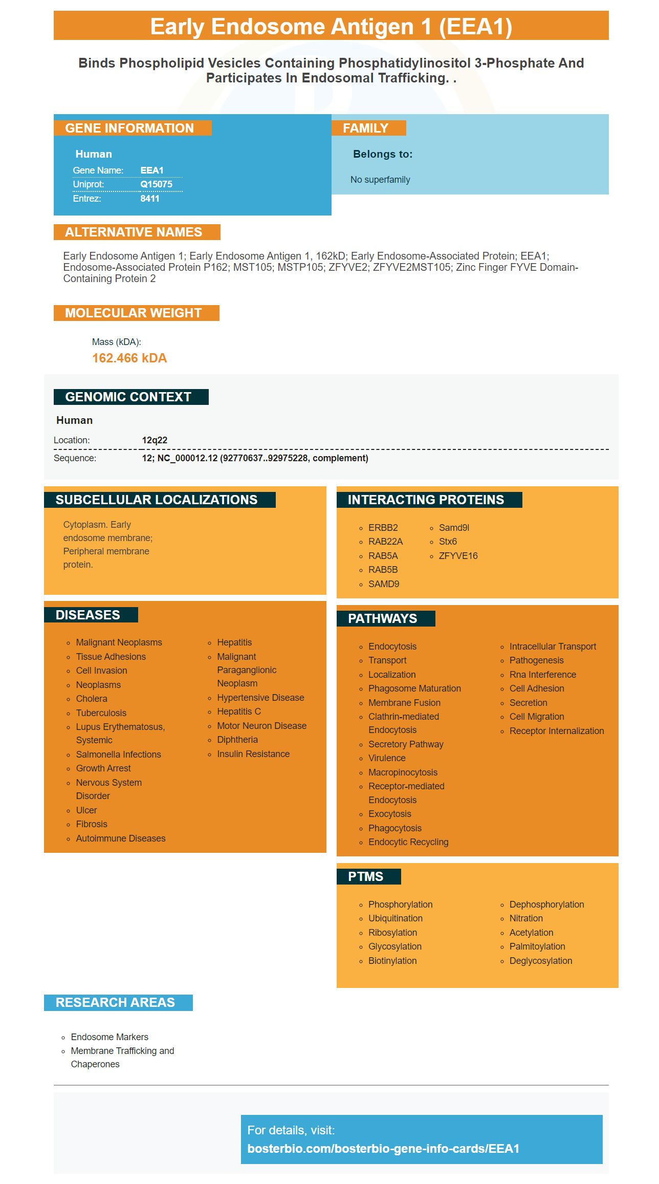
Human | |
---|---|
Gene Name: | EEA1 |
Uniprot: | Q15075 |
Entrez: | 8411 |
Belongs to: |
---|
No superfamily |
early endosome antigen 1; early endosome antigen 1, 162kD; early endosome-associated protein; EEA1; Endosome-associated protein p162; MST105; MSTP105; ZFYVE2; ZFYVE2MST105; Zinc finger FYVE domain-containing protein 2
Mass (kDA):
162.466 kDA
Human | |
---|---|
Location: | 12q22 |
Sequence: | 12; NC_000012.12 (92770637..92975228, complement) |
Cytoplasm. Early endosome membrane; Peripheral membrane protein.
This article will cover the most commonly used applications for the marker EEA1 (a membrane-bound Rab5 effector protein). It is also used as a co-stain in the APPL1. EEA1 is a useful marker in the study of CDE. Its application isn't limited to scientists in the U.S. but is applicable everywhere.
EEA1 is a membrane bound Rab5 effector protein. It docks the incoming Endocytic vesicles prior the joining with early endosomes. It is located on the subdomain that is responsible for early sorting endosomes and not on clathrin coated vesicles. Moreover, EEA1 is associated with filamentous material on the cytoplasmic endosomal domain, which is in line with its docking/tethering purpose.
We utilized antibodies that were specific for GST to identify the EEA1. BFA treatment did not affect the tubulation of EEA1, but did shift the distribution of EEA1 towards the pericentriolar compartment. More research is required to identify the molecular events that caused this change. The purified EEA1 was then detected by ELISA using rabbit anti-goat antibodies coupled to HRP.
The purified EEA1 was positioned on an SDS-PAGE Gel without boiling, prior to the test. Then, the purified EEA1 was incubated with soluble GST-Rab5 and GTPgS. The antibodies were then used to identify GST-Rab5. After incubating the EEA1 purified, they were placed on an affinity column with active GST-Rab5. The purified Rab5 was extracted from the beads using a glutathione elimination buffer consisting of 1mM GTPgS and 1% BSA.
The function of Rab GTPases is vital in the role of Rab5 effectors involved in membrane trafficking. These GTPases selectively interact with multivalent effector proteins to facilitate tethering, sorting, and fusion. This interaction between Rab5 and EEA1 is mediated by the C(2)H(2) zinc finger in the effectors.
Apart from their role in membrane fusion, Rabs also have intrinsic tethering activities. They recruit tethering factors to facilitate the formation of vesicles as well as expand their lumen. These tethering proteins might be involved in the determination of apical membrane initiation points. The role of Rab5 effectors is not clear but the presence of EEA1 will be of interest in future studies.
EEA1 was detected in both the postsynaptic as well presynaptic endosomes in the brain of a rat. Presynaptic cells also showed a decrease in EEA1, while MAP2 was found to be intact in synaptosomes. Rab5 is associated with both types of endosomes suggesting that it is involved in regulating endosomal trafficking.
EEA1 is a membrane-bound Rab5 effector that plays a vital function in the transport of endocytic vesicles. The union of early endosomes and endocytic vesicles allows for the redistribution of compounds the cytoplasm and into other parts of the cell. The FYVE domain's C-terminal targets early endsosomes , and docks with SNARES to facilitate membrane fusion.
EEA1 colocalises in an autophagy-competent line with ATG16L1 early endosomes. Bafilomycin inhibited lysosomal acidification and enhanced the ATG16L1/EEA1 colocalization. This suggests that ATG16L1 can be recruited to EEA1+ endosomes following chemical damage to the endomembrane. However, in autophagy-incompetent cells, this recruitment is reduced. In turn, compensatory mechanisms can affect the maturation of endosomes and biogenesis.
The analysis of the immunohistochemistry of EEA1 revealed that a specific antibody to the protein binds only early endosomal vesicles, including mCherry Vesicles. This shows that EEA1 may be used to identify the purity of the cytosolic portion.
The binding of receptors to plasma membrane is the endocytic pathway. The RTK is internalized and transported into the cell through clathrin-mediated endocytosis, allowing the cell to acquire cargo from the environment. Later, the vesicles join with the first endosomes which are homogeneous and contain receptors.
Additionally, EGFR is also a autophagy target, which targets organelles with damaged. The ablation of autophagy's essential proteins decreases EGFR recycling into the plasma membrane, thereby affecting the survival of cells and their homeostasis. Moreover, ablation of EEA1 hinders EGFR trafficking and endocytosis. Early endosomes have significant amounts of EGF.
EEA1 is an peptide that facilitates membrane invagination through its interactions with the PI(4,5)P2 structure. The N-terminal residues of the protein fold into an amphipathic Helix that inserts into the bilayer's outer leaflet and can bend the membrane. Other proteins can also form amphipathic-helices.
EEA1 regulates Rab5 and is essential in early endocytosis. Furthermore, it attracts effectors to early endosomes and aids in endosomal trafficking. Its ubiquitination-activated state promotes endosomal retention of repurposed transporters. Rab5 also acts as a mediator in endosomal membrane recycling.
The APPL1 marker is unique to the world of APPL endosomes. It interacts with several membrane receptors and signalling proteins, and plays multiple roles in cell growth and survival as well as metabolism. It also regulates b-catenin-dependent transcription and the remodeling of chromatin. The APPL1 gene has been involved in multiple signaling pathways, including cell proliferation and Apoptosis.
Abnova and GE Healthcare obtained purified GST-tagged APPL1. It was then immobilized onto glutathione-sepharose beads that contain 20 percent of glycerol. Cells were then permeabilized in a buffer containing 20 mM Tris-HCl pH 7.9, 1 mM EDTA, 5 MgCl2, 0.2 mM phenylmethylsulphonyl fluoride, and 0.1 M NaCl.
MG132 treatment of HeLa cells with APPL1 or vimentin showed that APPL1 as well as vimentin co-stain. MG132 treatment also reduced aggresome-related cell growth in co-treated cells as well as those in control. These results were different from those observed with MG132 by itself. Further studies are required to confirm the importance of co-staining APPL1 with EEA1 in vitro.
In vivo, APPL1 knockout mice showed impaired retrograde trafficking and the accumulation of peri-nuclear EGF. They also showed diminished the peri-nuclear storage of EGF and were extremely responsive to EGF. The results showed the direct connection between EGF and endosomes containing APPL1, which is a result of both APPL1 knockout mice as well as their wild-type counterparts.
APPL1 has many functions and is involved in a variety of cellular processes that include growth, proliferation, and survival. It is also closely linked to signal transduction and membrane traffic. It is involved in a variety of illnesses including cancer. An abnormality in the ubiquitin proteasome system results in the an accumulation of ubiquitinated substances, which accumulate as large inclusion bodies.
Proteasome inhibitors, such as MG132, block the proper localization of APPL1 in the endosomes. They also inhibit the APPL1 marker protein. This inhibitor reduces the number that APPL1 is present in endosomes by dramatically reducing the total intensity (the total of all vesicles in a cell)
Recent studies have proven that EEA1 could be an important marker in the development of CDE. This is a genetic disorder that affects endosomal systems. EEA1 is linked to endosomal sorting vacuoles. This finding is supported by immunofluorescent analysis of cells treated with BFA. Furthermore, the expression of EEA1 in these cells was reduced in the presence of BFA.
The lack of the globular ATP binding head domain and actin-binding domain of EEA1 suggests that EEA1 has a close evolutionary relationship with myosins from other species. The hydrophobic interactions between calmodulin and the amino acid phenylalanine which is necessary for calcium-to-neurmodulin bind and neuromodulin binding, is also disrupted due to the amino acid phenylalanine. This could be a significant connection in the context that the EEA1 gene is a significant indicator of CDE.
The heavy labeling of EEA1 could be associated with intracellular membranes and tubular or vesicular profile. EEA1 labeling is not compatible with tannic acid staining and HRP labeling. However, HRP-labeled vesicles were similarly labeled, which suggests that EEA1 is involved in transporting clathrin-coated vesicles from endosomes.
We discovered that p97 is responsible for the interaction of EEA1 and endosomes inside CDE cells. This model enabled us to detect the co-localization between EEA1 in COS7 cells and p97. It is interesting to note that p97 did not interfere with the membrane interaction of EEA1. Similarly, membrane interaction between EEA1-p97 was not required by p97.
EEA1 localizes to early endosomes, and the function it plays is not understood fully. Recent research suggests that it is present in greater quantities in the cytosol that was previously thought. This suggests that the EEA1 proteins can be easily dissociated from membranes. Two-dimensional immunoblots have shown that EEA1 also has an acidic pI.
We discovered that the EEA1's C2H2 domain is vital in its connection to the p97. Additionally, we discovered that p97 hinders EEA1's interaction with endosomes. Our findings suggest that EEA1 is a crucial marker for CDE. Although p97 plays an important role in CDE, its absence from endosomes inhibits the connection between p97 and EEA1.
PMID: 7768953 by Mu F.-T., et al. EEA1, an early endosome-associated protein. EEA1 is a conserved alpha-helical peripheral membrane protein flanked by cysteine 'fingers' and contains a calmodulin-binding IQ motif.
PMID: 9697774 by Simonsen A., et al. EEA1 links PI(3)K function to Rab5 regulation of endosome fusion.
*More publications can be found for each product on its corresponding product page