This website uses cookies to ensure you get the best experience on our website.
- Table of Contents
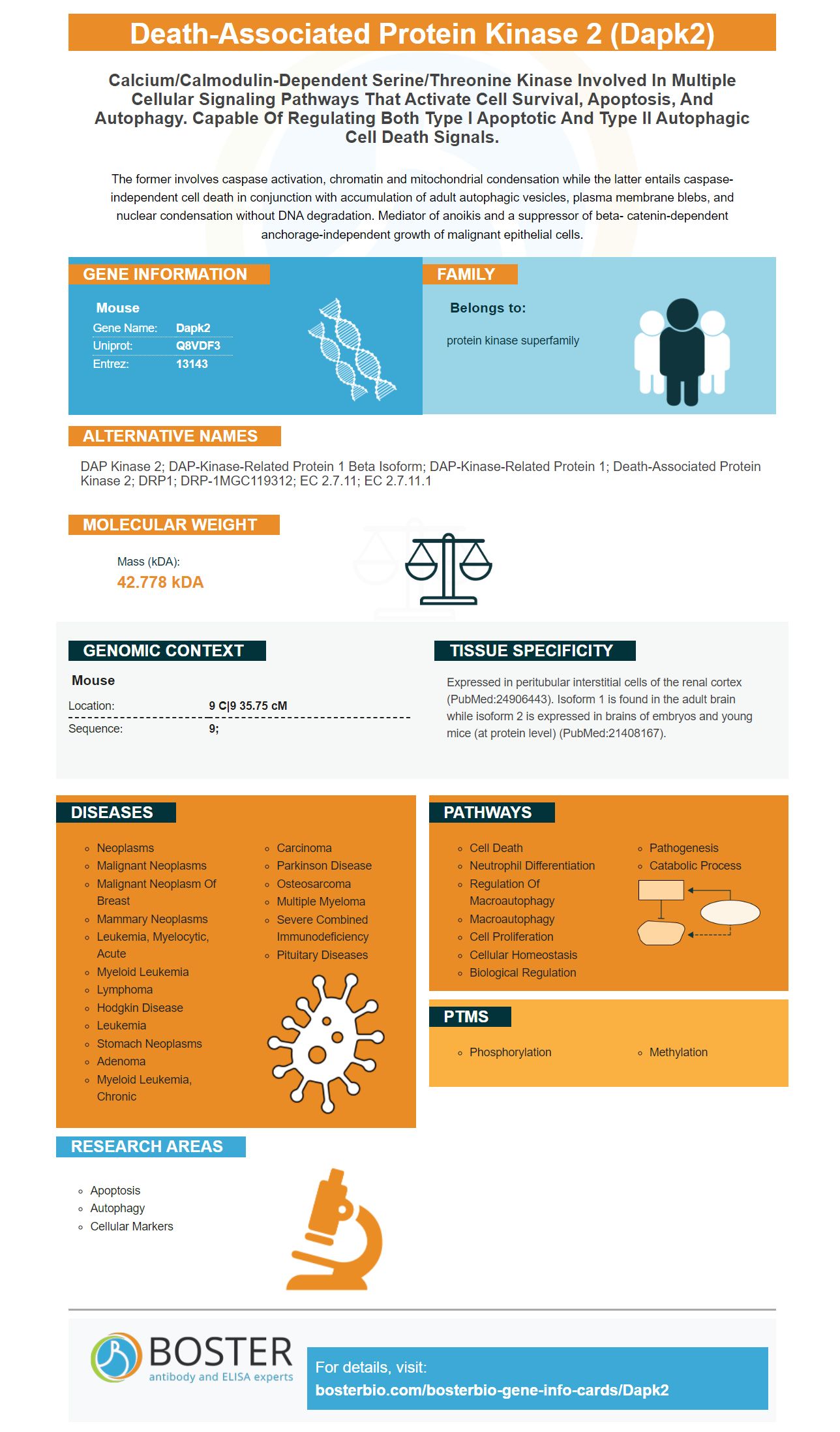
Facts about Death-associated protein kinase 2.
The former involves caspase activation, chromatin and mitochondrial condensation while the latter entails caspase-independent cell death in conjunction with accumulation of adult autophagic vesicles, plasma membrane blebs, and nuclear condensation without DNA degradation. Mediator of anoikis and a suppressor of beta- catenin-dependent anchorage-independent growth of malignant epithelial cells.
Mouse | |
---|---|
Gene Name: | Dapk2 |
Uniprot: | Q8VDF3 |
Entrez: | 13143 |
Belongs to: |
---|
protein kinase superfamily |
DAP kinase 2; DAP-kinase-related protein 1 beta isoform; DAP-kinase-related protein 1; death-associated protein kinase 2; DRP1; DRP-1MGC119312; EC 2.7.11; EC 2.7.11.1
Mass (kDA):
42.778 kDA
Mouse | |
---|---|
Location: | 9 C|9 35.75 cM |
Sequence: | 9; |
Expressed in peritubular interstitial cells of the renal cortex (PubMed:24906443). Isoform 1 is found in the adult brain while isoform 2 is expressed in brains of embryos and young mice (at protein level) (PubMed:21408167).
Boster Bio's DAPK2 marker is a good choice if you are looking for anti-DAPK2 antibodies. Did you know that this biomarker has a lot more versatility than you might think? For more information on its Specificity, Applications and Validation, read on. We also discuss the most common pitfalls and how they can be avoided when using biomarkers.
The Anti-DAPK2 Marker from Boster Bio was developed to detect a protein that regulates the signal pathways leading to type I apoptotic and type II autophagic cell death. Numerous biological assays used antibodies that detect Dapk2 when identifying the protein. The antibodies can be monoclonal (or polyclonal) and have been validated against panels containing 250 tissues. They are also quantitatively validated against known concentrations of recombinant proteins and untransfected cell lines to ensure high specificity.
DAPKs are regulated by non-coding RNAs, and it is important to identify these RNAs before studying the gene. DAPK2 regulates a wide range of genes, including Akt and protein kinase C (PKC). Interestingly, DAPK2 regulates activity of the CK/CRB2 and ERK1/2 subunits. These non-coding RNAs have been found to play key roles in regulating the expression of DAPKs in cancer.
DAPK2 is expressed at various developmental stages including embryonic and postnatal. It is absent from E15.5 and P0. DAPK2 expression at P14 is low, but still detectable within interstitial tissues. The accompanying image contains more information. (A & B are in gray respectively).
The DAPK2 genes is expressed in interstitial cell populations of the cortex. The Dapk2 nil mouse has normal kidney function in steady-state conditions. It is resistant to extracellular fibrosis and can withstand matrix deposition in experimental renal fibrillation. These results suggest that DAPK2 plays some role in kidney injury. It is important to note that this gene is expressed by many cell types, including fibroblasts, vascular endothelial cells, and muscle cells.
Ca2+/calmodulin regulates DAPK2's gene. The kinase-domain is 80% homologous with DAPK1, but the autoinhibitory sphere is only 20%. The autoinhibitorydomain is thought regulate DAPK2 activitiy by binding with Ca2+/CaM. The kinase domain and the CBD are structurally similar. These regions share the same regulatory mechanisms.
Four residues in the DAPK2 gene are phosphorylated in vivo. These include S367STS370's 14-3-3 binding molecular motif and two additional autophosphorylation points. The 14-3-3 binding site may play a role in DAPK2 activity and binding with 14-3-3 phosphorylated proteins. Furthermore, the C terminus of DAPK2 is phosphorylated in vivo.
The DAPK2 gene belongs to the family of death-associated proteins kinase genes. It is a multifunctional proteinkinase that regulates many processes in the body. DAPKs genes are involved in apoptosis and granulocyte differentiate, as well as motility regulation. Because of its role in many processes, DAPK2 is a potential target for anti-inflammatory therapies.
The use of a DAPK2 marker for cancer diagnostics has recently gained in popularity. It is a powerful and highly specific gene expression marker, and its use has become routine for researchers. Some researchers aren't happy with the current level of specificity. Many people aren't sure if the marker works. We will discuss the specifics of this marker. We will also give you a brief overview regarding the marker's main features.
The DAPK2 marker phosphorylates DAPK2. The antibody reacts with phosphorylated DAPK2 Ser289, and is able to detect this phosphorylation even in the presence AMPK. This marker only gave strong signals when incubated in DAPK2K42A and not in the double mutant S289A. Ser289 is a conserved amino acid located in a short loop between DAPK2's CaM-binding domain and its kinase domain.
The DAPK2 kinase is regulated by phosphorylation and 14-3-3 binding, respectively. AKT has been proposed to be a candidate for this inhibition of DAPK2. We have also shown that S289D, S308D and S308D mutants cause blebbing in 45% GFP-positive cells. We also demonstrated that the S289D mutation provides a novel mechanism for upregulating DAPK2 activity that is independent of CaM binding and Ser308 dephosphorylation.
DAPK1 methylation is an important biomarker to detect gastrointestinal cancers early. Aberrant methylation is associated with gastric cancer tumorigenesis. Although little is known about the role it plays in cutaneous carcinoma, it may be a target for new treatments. This information can help doctors plan for successful treatment.
DAPK2 is a member of the DAPK family of proteins. The domains occupied DAPK2 family members reflect the location and function of DAPK2's gene markers. This means that the DAPK2 gene expression marker is highly specific to this protein. DAPK2 methylation for breast cancer is not specific enough to be specific for it.
A variety of biological tests can use the DAPK2 indicator. These antibodies can be monoclonal or multiclonal and react with DAPK2 on a variety of animal samples. Boster Bio creates antibodies against the marker DAPK2 in rabbits or mice. The DAPK2 gene encodes a protein that regulates type I apoptotic and type II autophagic cell death signals.
The DAPK2 gene is involved in the development and maintenance of bone growth and metabolism. In addition to its role in bone metabolism, DAPK2 promotes osteogenesis, which can be useful in bone development. There are many diseases that can be caused by DAPK1 exclusion. It is important to learn how DAPK1 can also be suppressed, and how it affects bone health.
PMID: 10376525 by Kawai T., et al. Death-associated protein kinase 2 is a new calcium/calmodulin- dependent protein kinase that signals apoptosis through its catalytic activity.
PMID: 10629061 by Inbal B., et al. Death-associated protein kinase-related protein 1, a novel serine/threonine kinase involved in apoptosis.