This website uses cookies to ensure you get the best experience on our website.
- Table of Contents
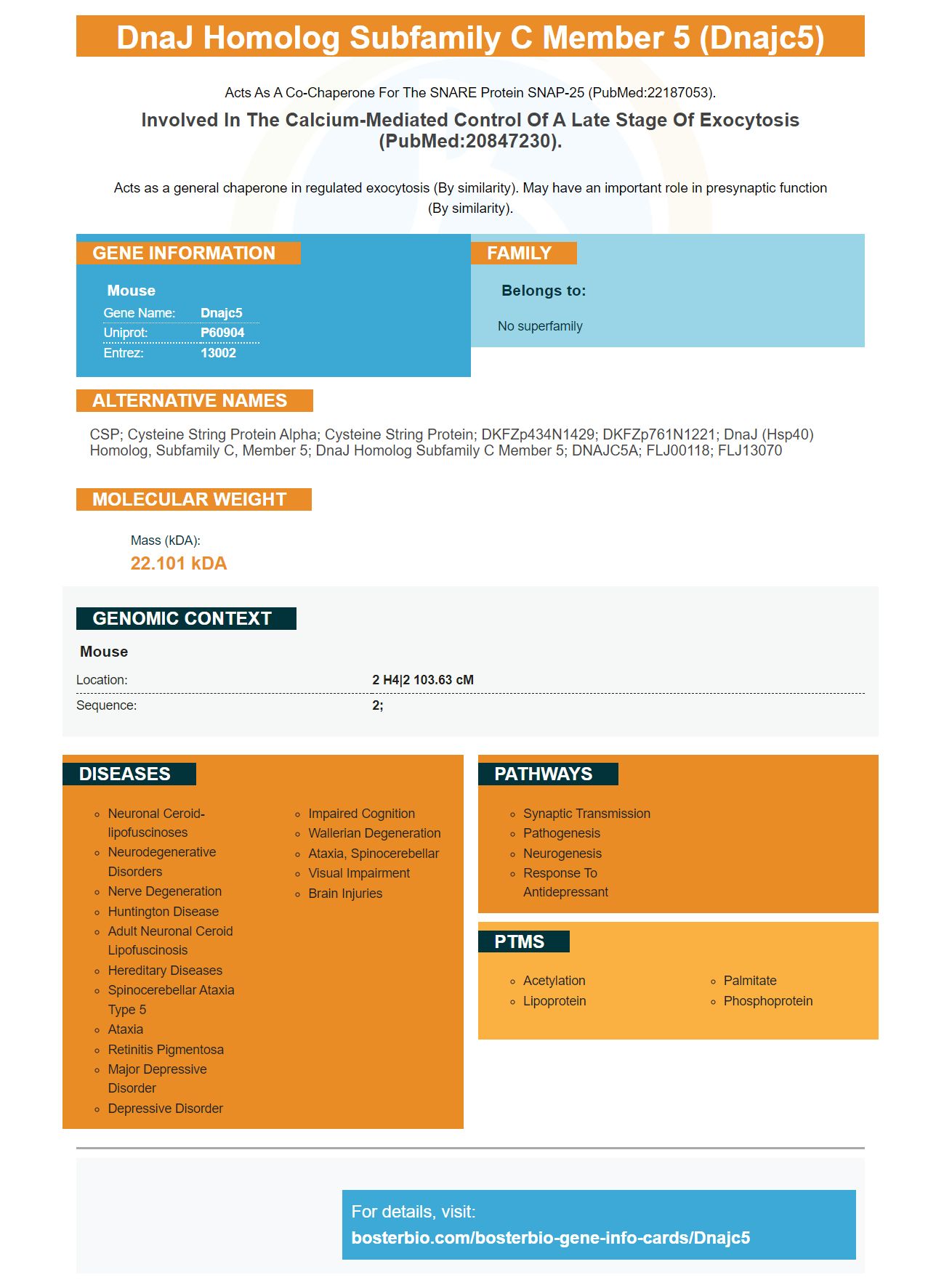
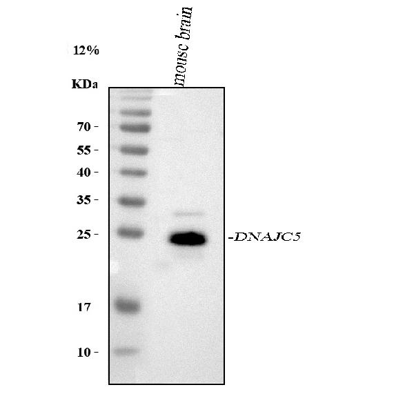
Facts about DnaJ homolog subfamily C member 5.
Acts as a co-chaperone for the SNARE protein SNAP-25 (PubMed:22187053).
Involved in the calcium-mediated control of a late stage of exocytosis (PubMed:20847230).Acts as a general chaperone in regulated exocytosis (By similarity). May have an important role in presynaptic function (By similarity).
Mouse | |
---|---|
Gene Name: | Dnajc5 |
Uniprot: | P60904 |
Entrez: | 13002 |
Belongs to: |
---|
No superfamily |
CSP; cysteine string protein alpha; Cysteine string protein; DKFZp434N1429; DKFZp761N1221; DnaJ (Hsp40) homolog, subfamily C, member 5; dnaJ homolog subfamily C member 5; DNAJC5A; FLJ00118; FLJ13070
Mass (kDA):
22.101 kDA
Mouse | |
---|---|
Location: | 2 H4|2 103.63 cM |
Sequence: | 2; |
The DNAJC5 Marker can be used for many purposes, including detection of cancer cells. The marker can be detected using two methods, ELISA and autoradiography. This article describes the Detection techniques, Protein transfer efficiency, as well as Autoradiography film. This guide will prove invaluable to all DNAJC5 users. It will also help you choose the best test for your application.
A DNAJC5 region is located in the genome of a person suffering from ANCL (adult-non-white blood cells) leukemia. This region was amplified using PCR, and the product was sequenced using a Dye Terminator cycle sequencing kit available from Applied Biosystems. The PCR products were sequenced within a frame using an ABI3500XL Avant Genetic Analyzer. Sequencing Analysis software was used to analyze the data. Direct sequencing of genomic DNA fragments was also used to identify candidate mutations. Sequencing analysis of DNAJC5 gene fragments was conducted with primers listed in Table S1.
We found that overexpression of the gene can stimulate MAPS activity by recruiting MAPS sub-substances to the membrane in a study with recombinant DNAJC5. Biochemical fractionation demonstrated that recombinant DNAJC5 bound to membranes. The binding was saturable and showed that there may be a limiting element that regulates membrane-binding. However, membrane-associated Flag-GFP1-10 did not co-localize with endogenous DNAJC5 in the peri-nuclear compartment, which corresponds to lysosomes.
After transfection, COS7 cell lines were used to detect the DNAJC5, a protein that is found in late endosomes or lysosomes. After incubation, live-cell imaging showed that DNAJC5-transfected CoS7 cells had mCh–GFP1-10. This rapidly diffuses in the cytosol. Photobleaching however reduced the signal of mCh-GFP1-10, which allowed visualization of trapped endosomes.
ExoU is an essential pore-forming toxin for cytotoxicity in human epithelial and other cells. The PA14 virus causes cytotoxicity. DNAJC5 and ExoU are required. DNAJC5 is necessary for the PA14 toxin's necrosis activity. DNAJC5 is required for ExoU toxicity of host cells. ExoU mutations inhibit this activity.
Both the identification and detection of DNAJC5 are based on inner codification analysis. To determine the canonical DNAJC5 image, the first step of candidate detection is to apply perspective transform to it. After the image is obtained it is thresholded by Otsu to separate out the black and/or white bits. The size of the marker is used to divide the image into cells. Bits are either black or white based on the number of pixels that fall within each cell.
Debugging is the next step for detecting DNAJC5 marker. The aruco module detects markers. It uses its detectMarkers() function in order to determine if the mark is present. DetectionMarkers(), the most important function in a module, is it? Once a mark has been detected, the module's other functions are dependent on it. The markerCorners parameter lists the corners of all detected markers. Each corner starts at the top.
Sample preparation is crucial for clear Western blots. Sample preparation is crucial. The right method of sample preparation can make all the difference between a plain blot or one that looks beautiful. Boster offers many lysis buffers to meet your needs. Use the guide to help you choose the best extraction kit. Different types require different equipment. They also require different speeds and efficiency. Your needs and desired results will determine the best choice.
Traditionally, wet transfer is the most common method. This method is very efficient but takes a lot more time and effort. Life Technologies' iBlot2r systems are semi-dry blotters. Both methods require some buffer for transfer, but they work faster than wet transfer. Trans-Blot Turbo marker by Bio-Rad and Boster Bio DNAJC5 marker increases protein transfer efficiency.
DNAJC5 is a widely used gene expression marker for human cancer. Its sensitivity can be improved by the use of Picokine(tm) ELISA kits. These kits have been validated against more than 250 human tissues. It is possible to determine the antibody's high affinity by testing it against untransfected cell lines containing known amounts of recombinant protein.
PMID: 17034881 by Boal F., et al. Cysteine-string protein isoform beta (Cspbeta) is targeted to the trans-Golgi network as a non-palmitoylated CSP in clonal beta-cells.
PMID: 20847230 by Boal F., et al. A charged prominence in the linker domain of the cysteine-string protein Cspalpha mediates its regulated interaction with the calcium sensor synaptotagmin 9 during exocytosis.