This website uses cookies to ensure you get the best experience on our website.
- Table of Contents
15 Q&As
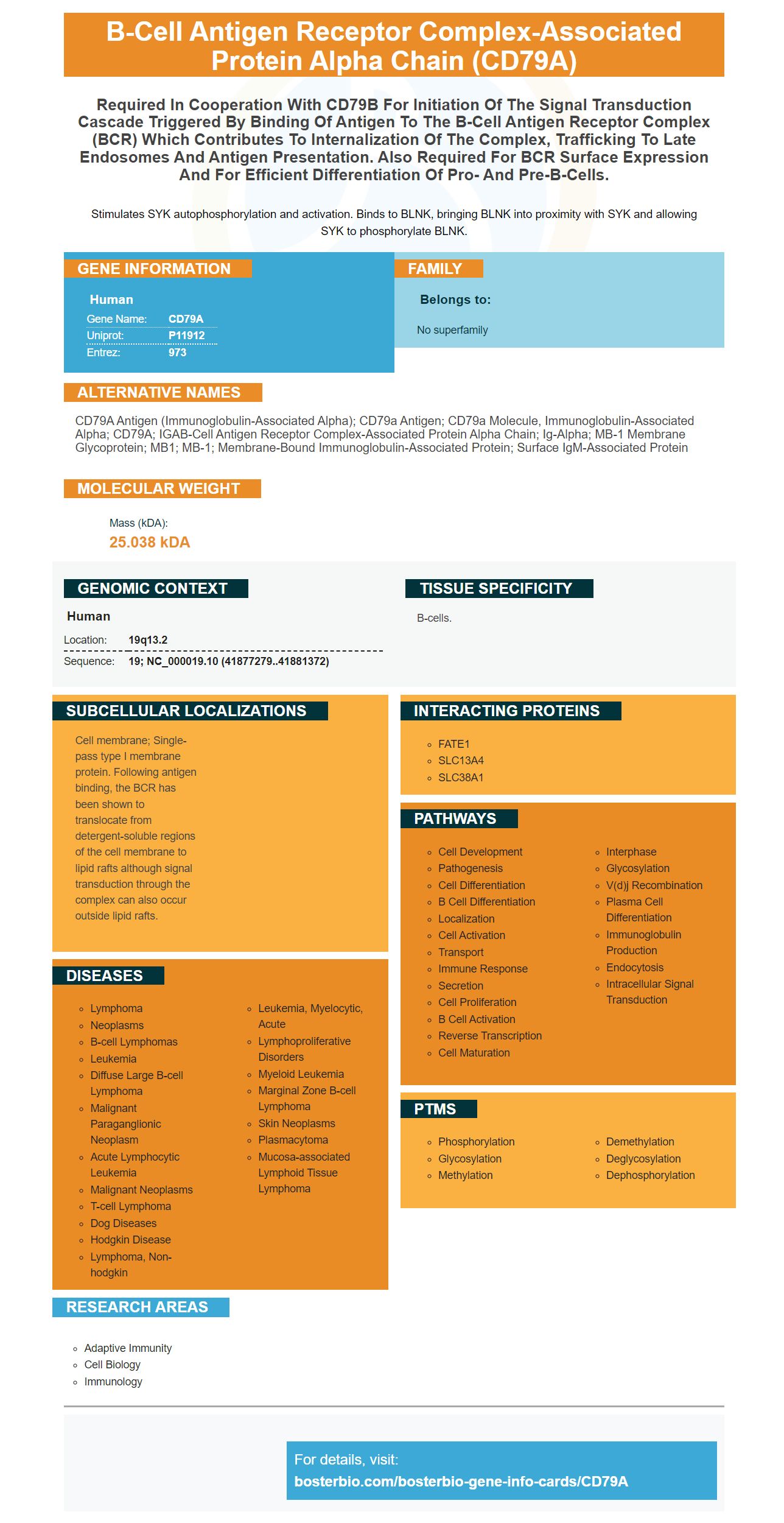
Facts about B-cell antigen receptor complex-associated protein alpha chain.
Stimulates SYK autophosphorylation and activation. Binds to BLNK, bringing BLNK into proximity with SYK and allowing SYK to phosphorylate BLNK.
Human | |
---|---|
Gene Name: | CD79A |
Uniprot: | P11912 |
Entrez: | 973 |
Belongs to: |
---|
No superfamily |
CD79A antigen (immunoglobulin-associated alpha); CD79a antigen; CD79a molecule, immunoglobulin-associated alpha; CD79A; IGAB-cell antigen receptor complex-associated protein alpha chain; ig-alpha; MB-1 membrane glycoprotein; MB1; MB-1; Membrane-bound immunoglobulin-associated protein; Surface IgM-associated protein
Mass (kDA):
25.038 kDA
Human | |
---|---|
Location: | 19q13.2 |
Sequence: | 19; NC_000019.10 (41877279..41881372) |
B-cells.
Cell membrane; Single-pass type I membrane protein. Following antigen binding, the BCR has been shown to translocate from detergent-soluble regions of the cell membrane to lipid rafts although signal transduction through the complex can also occur outside lipid rafts.
If you've ever wondered how to use Boster Bio, HM47, Picokine, or Picoband in your cancer research this article is designed for you. We'll guide you through each product and give you an overview of their distinctive features and best uses. We'll also go over their history, the benefits and how they can help you with your research.
Pre-B-ALL is a condition in which tumors that express CD79a are not positive for other B cell markers like CD20 and the T-cell receptor CD45RA. There are a variety of applications for CD79A antibodies. These antibodies can be used to identify B-cell neoplasms and in patients who have received a deficient CD20 antibody treatment. This article will examine the molecular biology and use of CD79a/CD79b in T-cell and B-cell tumors.
The monoclonal antibody CD79A recognizes the mb-1 peptide that is a B-cell antigen receptor compound. This antibody is superior to other markers available commercially of B-cells. There are many different applications of this marker. Here are a few of them. They comprise the following. The HM47 antibody reacts with mouse, human as well as rat, dog and monkey, in addition to macaques, primates, horse, chicken, and rabbit.
CD79A is found in a range of B-cell neoplasms. In contrast, it is found in only 50 percent of plasma cell tumors. In addition, CD79a is found in L&H cells in nodular lymphocyte-predominant Hodgkin lymphomas. About 20% of cases have high levels of CD79A positivity. Certain myeloid leukemias have CD79a.
The CD79A marker is highly specific in defining the B-cell lineage. It is considered equivalent to CD3 by a variety of authors. Its high degree of specificity in the identification of B-cells has led to its selection by the European Group for Immunological Characterization of Acute Leukemias as a powerful marker of B-cell differentiation. In addition, since it is found in all species, it can be useful in the identification of B-cell lymphomas that are not human.
The CD79A, CD79b components of this protein are almost exclusively expressed on B cells and B-cell Neoplasms. The markers are expressed prior the rearrangement of the immunoglobulin heavy chain gene and disappear later than CD20 during the late stages of B-cell differentiation. These proteins are employed for the diagnosis of T-cell and B-cell neoplasms.
The CD79A marker is a protein on the cell surface that is extremely specific for B-cell differentiation is known as a CD79A marker. According to the EuroFlow Consortium, CD79a is more sensitive and specific than CD22 which makes it an excellent pan cell marker to assess B-lineage. Additionally, CD22 is not lineage-specific and is expressed in high levels on mast cells, normal basophils and dendritic cells.
The CD79A marker can also be used for diagnosing B-ALL patients. Flow cytometry analysis can be difficult to obtain particularly in developing countries. Furthermore, the number of available markers for lineage classification is limited, which may cause an inaccurate diagnosis. The CD79A marker is a good choice for a variety of reasons, including to study the morphology of B-cells and the bone Marrow Trephines.
The CD79A marker is a biomarker that is highly specific for B-cell lymphomas. It binds to an antigen in B-cells, and is known as mb-1. It is a membrane protein , with an extracellular immunoglobulin region and a small cytoplasmic region. CD79A's gene is located at 19q13. The gene is expressed throughout the development in all B cells. It is expressed in B-lineage lymphomas, as well as non-B cells. In this way the CD79A marker is a vital indicator in the diagnosis of B-cell lymphomas.
The Boster Bio Anti-Cd79A antibody Picoband reacts with Rat and Mouse. The antibody can be stored at -20°C for up to a year. The solution contains Na2HPO4 (trehalose) Blocking peptides can be purchased at an additional cost based on the length of the immunogen. Picoband and CD79A markers are complementary tools for precise B-cell characterization.
The CD79A marker is extensively used in immunological assays in different fields. There are many monoclonal as and polyclonal antibody that react to CD79A in various animal samples. Boster Bio has developed CD79A antibody that detects CD79A in B cells. This lets researchers determine whether samples are positive or not for the CD79A protein. The antibodies are able to detect the CD79A protein and are essential to determine BCR surface expression as well as for efficient differentiation of pre and proB cells.
Boster Bio produces ELISA kits and research antibodies for cancer, neuroscience inflammation, developmental biology. They offer a broad range of antibodies that have been tested against an array of 250 tissue samples. The antibodies can be quantified against the known amounts recombinant proteins and untransfected cell line levels. Boster Bio also offers technical support to back their products. They are available to answer any questions you may have.
PMID: 1395095 by Leduc I., et al. Structure and expression of the mb-1 transcript in human lymphoid cells.
PMID: 1534761 by Mueller B.S., et al. Cloning and sequencing of the cDNA encoding the human homologue of the murine immunoglobulin-associated protein B29.