This website uses cookies to ensure you get the best experience on our website.
- Table of Contents
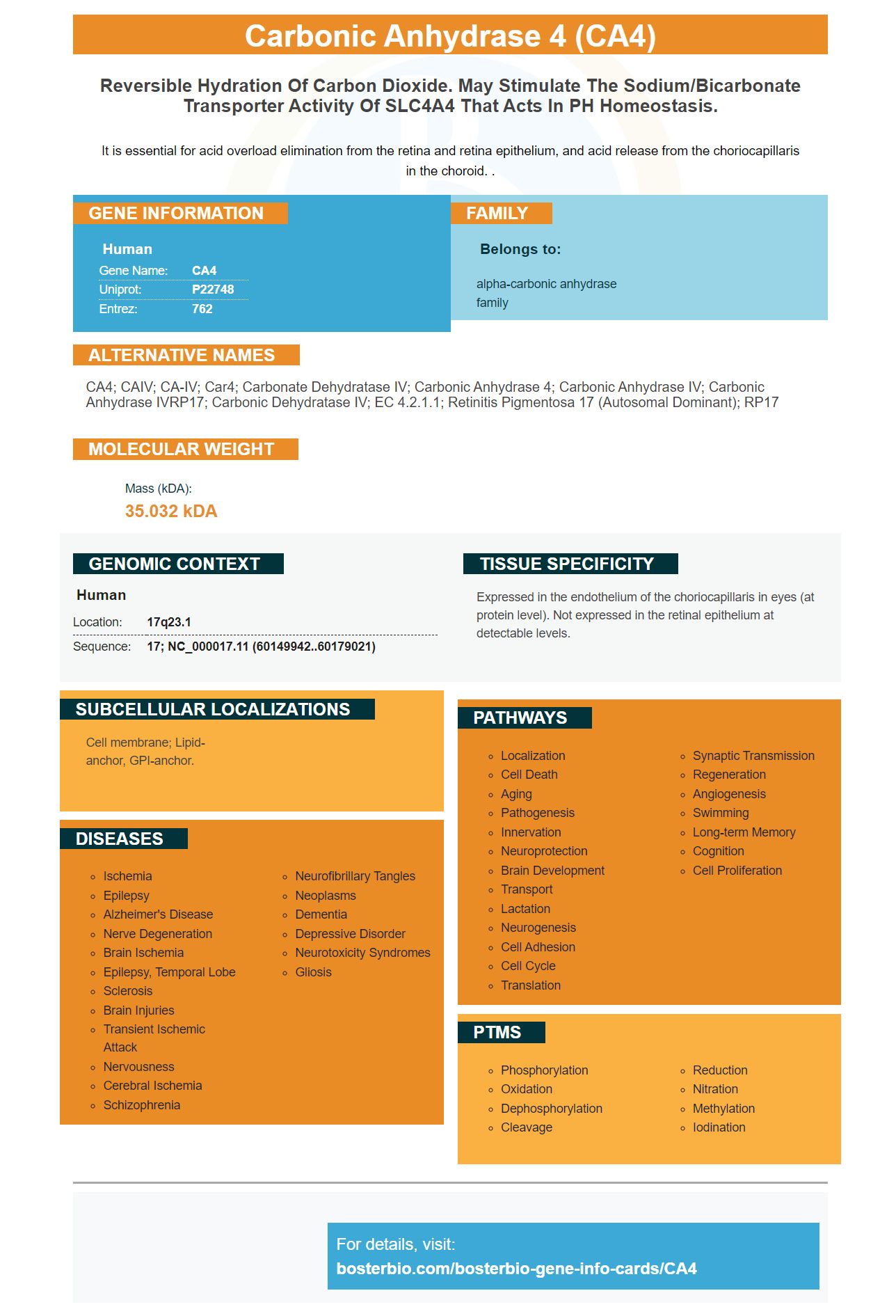
Facts about Carbonic anhydrase 4.
It is essential for acid overload elimination from the retina and retina epithelium, and acid release from the choriocapillaris in the choroid. .
Human | |
---|---|
Gene Name: | CA4 |
Uniprot: | P22748 |
Entrez: | 762 |
Belongs to: |
---|
alpha-carbonic anhydrase family |
CA4; CAIV; CA-IV; Car4; Carbonate dehydratase IV; carbonic anhydrase 4; Carbonic Anhydrase IV; carbonic anhydrase IVRP17; carbonic dehydratase IV; EC 4.2.1.1; retinitis pigmentosa 17 (autosomal dominant); RP17
Mass (kDA):
35.032 kDA
Human | |
---|---|
Location: | 17q23.1 |
Sequence: | 17; NC_000017.11 (60149942..60179021) |
Expressed in the endothelium of the choriocapillaris in eyes (at protein level). Not expressed in the retinal epithelium at detectable levels.
Cell membrane; Lipid-anchor, GPI-anchor.
The best way to use the CA4 marker in your research is to test its protein level in SO-RB50 cells. Boster Bio, the company Boster Bio makes monoclonal antibodies for this marker. These antibodies have high affinity and are incredibly sensitive. You can purchase your CA4 marker monoclonal antibody today. Boster Bio also offers a vast array of services to researchers and scientists. You can also avail technical materials for free and 24-hour customer support.
The anti-adiponectin antibodies of mice recognize human Adiponectin. Also called Acrp30 or adipocyte-complement related proteins. It is an amino acid adipokine of 244 amino acids that regulates glucose metabolism as well as catabolism of fatty acids. It circulates in the form of a polymer with different molecular weights, including trimers, hexamers, and Oligomers.
The purified mAbs recognized the adiponectin forms in human serum to different levels. KH7-41 recognized both MMW and HMW isoforms in rat and mouse serum. KH4-8 recognized the MMW isoform, while KH7-41 was not specific. There are antibodies available for both MMW- and HMW.
The cDNA that contained mature rabbit adiponectin was amplified using an PCR procedure and then placed into a pGEM-T vector using the T/A cloning method. The protein sequence that resulted was the adiponectin protein and had a concentration of 0.29 +/-0.02 mg/ml.
Boster Bio's Monoclonal Antibody recognizes adiponectin's molecular weight. This protein has anti-inflammatory, anti-arthritic and inhibits the inflammation response. It is also utilized in rodent models and human synoviocytes that resemble fibroblasts. So, it may have therapeutic potential in the near future.
In mice, treatment with Ad-APN inhibited the formation of 30 percent of lesion. In humans Adiponectin levels were low. were linked to the progression of subclinical coronary atherosclerosis. These results support the idea that Ad-APN plays an important role in atherosclerosis. Adenovirus infection causes the liver to produce Adiponectin. It is found in adipocytes, fibroblasts and other tissues.
In mice, adiponectin suppressed the expression of pro-inflammatory cytokines, and reduced joint degradation in CIA mice. It also increased the amount of adiponectin that was found in joint fluid of patients suffering from RA. It could play a significant role in joint inflammation. Adiponectin-mAbs did not decrease inflammation markers.
To determine the impact of adiponectin on the adipocyte's migration, this new antibody was used to detect adipocyte migration. In addition, it inhibited movement of JAR and HTR-8 cells. It also has the potential to regulate MMP-2 activity. To test this hypothesis this antibody may be useful in human research.
Two human cell lines SORB50 and Y79 were used to study the effects of miR-483-3p on the expression of the ECM1 genes. Western Blots were used to determine the expression of ECM1 in both the Y79 and SO-RB50 cell line. We found that SORB50 cells had a higher apoptosis rate when we used the miR-4866-3p mimic.
The cell lines Y79 and Rb were placed in 6-well plates to determine the levels of protein of SO-RB50 cells. The Y79 cell line was grown in high-glucose Dulbecco's modified Eagle's medium that contained 15% bovine serum fetal. The cells were exposed various proteins in the media and then cultured in humidified atmosphere at 5% CO2.
We also performed Western blot experiments to determine the protein levels in Y79 and SO-RB50 cells. We found that miR-486-3p controlled Bcl-2 and Bax, while increasing the levels of Bax and C caspase-3 within SO-RB50 cells. These results suggest that miR-486-3p could serve as a biomarker for RB.
After the SO-RB50 cells were harvested, we used a bicinchoninic acid assay kit to determine the amount of protein in the supernatant. We eliminated any excess culture fluid after 48 hours. The infected cells were stained with 0.1% crystal violet in the presence of 44% paraformaldehyde. The supernatant was evaluated for protein concentration using a kit made by Thermo Fisher Scientific.
The overexpression of NRMT reduced apoptosis and increased cell viability and repressed the process of apoptosis in retinoblastoma cell. Furthermore silencing CENPA prevented the pro-proliferative effects of Myc and increased the sensitivity of cells to CDDP. These results demonstrate the importance of NRMT in detecting and assessing the levels of expression of RB protein.
The study identified B cells that react to an antigen that had low affinity. They were then tested for their affinity to HEL proteins HELWT and HEL3X. Serum anti-HEL IgG1 on day 15 of the two responses bound HELWT with high affinity and HEL3X with a near equal affinity. These results confirm affinity maturation.
Cells were fixed in PBS and then grown for 48 hours in darkness. Next, cells were stained with a murine anti-CD3–AF700, anti–CD4–BV421 or anti–CD8–BV711. The cells were then processed using the BD Fortessa X20 the cytometer to perform a flow cytometric analysis. The data were analysed using FlowJo(tm).
High-affinity antibodies are derived from a hybridoma and has a molecular mass of 23,635 Da. However, this type of antibody often contains unwanted light chains and has no reproducibility. It is important to select high-affinity primary antibodies using the CA4 marker. REAfinity antibodies are created in mammalian cells through a biologically-defined culture environment. They also undergo rigorous analytical processes.
L19 A high-affinity human antibody fragment that targets the EDB domain of fibronectin. In tumors, it effectively targeted tumor vessels. L19 is able to absorb 8.2% of its dose. The ratio of blood-to-tumor ratios were 1.9, 3.7, and 11.8 at three five, six, and 24 hours. L19's ability to target tumors was not dose-dependent within a range of 0.7 to 10 mg.
Moreover, mAbs can deliver drugs or toxins to specific areas. These antibodies are used in antimicrobial and cancer therapy applications. Covalent bonding is utilized to connect most mAbs with the immunoglobulin molecules. In contrast to other molecules, the covalent bonding between the drug and antibody stops premature dissociation. Although the first drug conjugates were constructed with different quantities of both antibody and drug but later techniques have improved their consistency and accuracy. The biomarker CA4 can be used to identify mAbs.
Apart from being able to recognize antigens, antibodies perform other functions. They aid in the detection and classification of antigens. They are often raised against a specific antigen and can be used in conjunction or as a single. In microscopy, they are typically coupled with enzymes or fluorescent proteins. They are commonly used in immunofluorescence research. This method is the most efficient to detect antigens.
Researchers are looking for an easy, low-cost assay. Schistosomiasis (the most prevalent neglected tropical disease is responsible for an estimated 2.6 million lost years of disability-adjusted life each year. The current treatment for schistosoma is PZQ that has a low sensitivity and is impeded by drug resistance. The Schistosoma mansoni genome was recently released, which has fueled research into parasite biology. It also opened the door to the identification of drug targets.
Colorimetric tests for the CA4 protein are possible with a biochemical marker. They can be used for viability assessment and to measure the metabolic activity of cells. They require optically clear walls and bottoms. These methods are more expensive than colorimetric dyes. They are limited in their application to non-adherent cells.
A rapid colorimetric assay based on the phosphatidylcholine-phospholipase C gene has been developed by researchers. Researchers have developed a rapid colorimetric test that can detect eight hundred micrograms of phospholipase C per mL (or 2.82 CFU/mL) 10 times more sensitive than the traditional PCR-based method. Furthermore, the test showed no evidence of cross-reactions with bacterial species that are closely related.
PMID: 1311094 by Okuyama T., et al. Human carbonic anhydrase IV: cDNA cloning, sequence comparison, and expression in COS cell membranes.
PMID: 8325641 by Okuyama T., et al. Genomic organization and localization of gene for human carbonic anhydrase IV to chromosome 17q.