This website uses cookies to ensure you get the best experience on our website.
- Table of Contents
1 Citations 5 Q&As
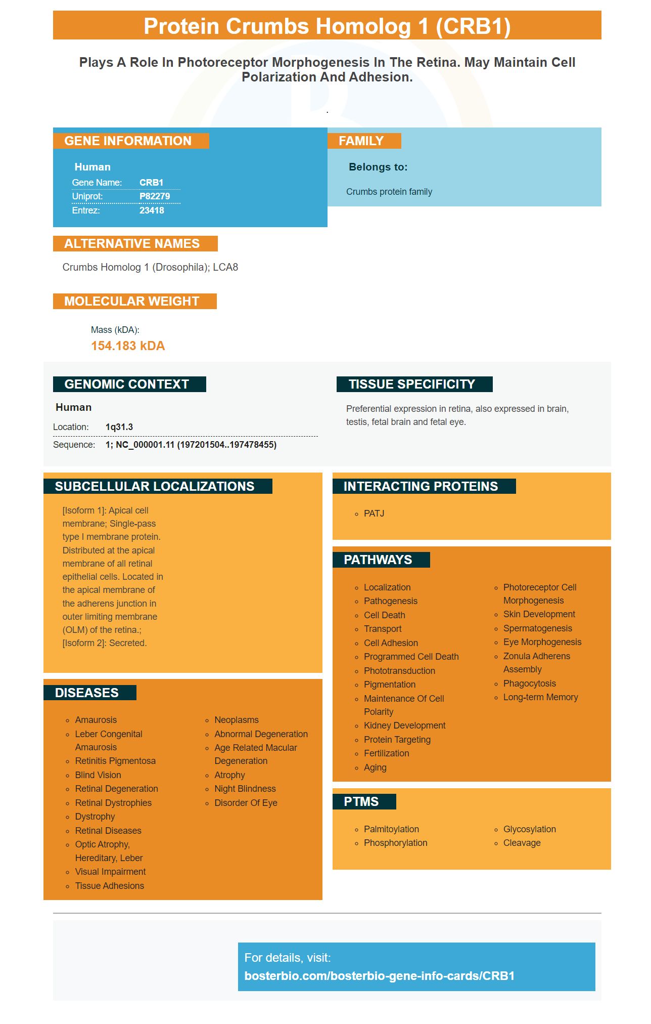
Facts about Protein crumbs homolog 1.
.
Human | |
---|---|
Gene Name: | CRB1 |
Uniprot: | P82279 |
Entrez: | 23418 |
Belongs to: |
---|
Crumbs protein family |
crumbs homolog 1 (Drosophila); LCA8
Mass (kDA):
154.183 kDA
Human | |
---|---|
Location: | 1q31.3 |
Sequence: | 1; NC_000001.11 (197201504..197478455) |
Preferential expression in retina, also expressed in brain, testis, fetal brain and fetal eye.
[Isoform 1]: Apical cell membrane; Single-pass type I membrane protein. Distributed at the apical membrane of all retinal epithelial cells. Located in the apical membrane of the adherens junction in outer limiting membrane (OLM) of the retina.; [Isoform 2]: Secreted.
Picoband Anti-CRB1 antibody from Boster Bio can be used to detect CRB1 in biological samples. Here are the advantages of this product and how you can apply it to your research. You can also submit results for species-specific samples and applications , and receive credit for the product. This product is accessible to all scientists across the world. The advantages of this product include:
Boster Bio Anti-CRB1 Antibodies are available in different formats like ELISA and Flow Cytometry. The antibodies are tested to identify specific proteins in samples of various formats. The antibodies can be utilized in a variety of tests since they are highly specific and high affinity. High quality antibodies from the company are featured in more than 29,000 scientific publications.
The CRB1 gene encodes the Crumbs homolog 1. It is akin to Drosophila's crumbs proteins. It is located in the stalks of the fly's photoreceptor. It may be a component of a molecular framework that regulates proper polarity development within the eye. This gene is related to severe forms of retinitis, and Leber congenital aurosis. There are many variants of the transcript in human and flies, that result in multiple CRB1 antibodies.
The PCR-based method of sequencing has many advantages over conventional DNA-based methods for finding the CRB1 gene. CRB1 contains two coding exons which each encode a distinct protein. Both are expressed in the brain and retina and may be involved in the normal development of photoreceptors. It is believed to be part of a larger system that determines the orientation and structure of these cells. The CRB1 marker can be detected by photoreceptors and in connections between cells.
A variety of cDNA sequences of Crb1 have been submitted to Genbank. Some of them are MT470365 (human CRB1-A), MT470366 (human CRB1-B), and MT470371-A2 (mouse Crb1-A2). Fig. 8d presents the results of three independent tests using the same antibodies. The Western blots represent the results of all three tests.
The screening of the CRB1 gene for mutations has led to results for 15 novel variants. These mutations are missense mutations, nonsense mutations, frameshift indels, as well as splicing-site mutations. The authors determined their pathogenicity through determining if they were co-segregating or not present in the 330 healthy Spanish control genomes. The early detection of LCA or EORP may be possible by detection of the CRB1 marker.
Studies of the molecular structure and function of CRBP1 markers provide icomrtant insights into the processes that lead to the formation of the retina. Researchers have found that CRBP1-A-B transmembrane protein has significant overlap with the Crb1-A however it differs in its intracellular domain. The CRB1-A gene is conserved. However, the Crb1B protein exhibits an unique in vivo conformation as predicted by lrCaptureSeq. Mice that lack the 5' exon do not express the protein in vivo and they don't have the intracellular domain.
The CRB1 marker, a protein that associates with adherent junctions which connect RMG cells to cone photoreceptors in the macula, is known as the CRB1. This is a phenotype known as MacTel 2 in humans. It is also linked to abnormal blood flow and is icolicated in a variety of processes. Detection of the CRB1 marker in vitro is helpful for determining the likelihood of developing macular degeneration.
The CRB1 gene has been extensively studied in both humans and mice. CRB1B, a human-specific gene, has five exons (5') and one exon (3'). Conventional short-read sequencing is not effective in distinguishing between these two exons. However, lrCaptureSeq can detect these variants. These studies have shown that CRB1 mutations can affect both the optic nerve and the macula.
The mutations that caused arRP and LCA were identified in the 404 families that had index cases. These families were later checked for rare CRB1 gene mutations using commercial genotyping chips built on APEX technology. Once the mutations were identified, they were confirmed by Sanger sequencing. PCR-based DNA sequencing confirmed the familial segregation. The study's findings revealed that the CRB1 gene is linked to the disease in four patients with Cys948Tyr and seven other missense mutations.
PMID: 10508521 by den Hollander A.I., et al. Mutations in a human homologue of Drosophila crumbs cause retinitis pigmentosa (RP12).
PMID: 11734541 by den Hollander A.I., et al. CRB1 has a cytoplasmic domain that is functionally conserved between human and Drosophila.
*More publications can be found for each product on its corresponding product page