This website uses cookies to ensure you get the best experience on our website.
- Table of Contents
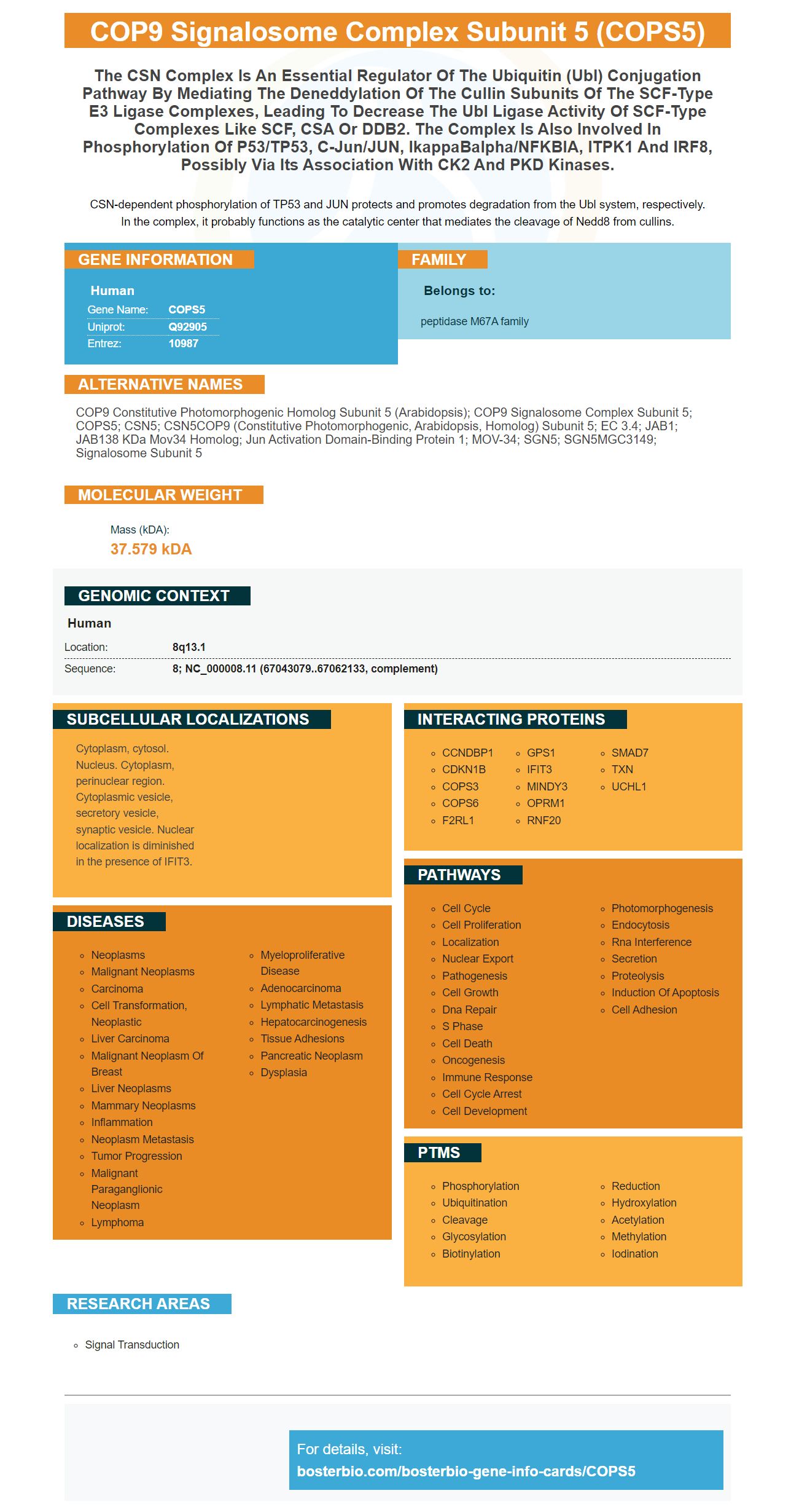
Facts about COP9 signalosome complex subunit 5.
CSN-dependent phosphorylation of TP53 and JUN protects and promotes degradation from the Ubl system, respectively. In the complex, it probably functions as the catalytic center that mediates the cleavage of Nedd8 from cullins.
Human | |
---|---|
Gene Name: | COPS5 |
Uniprot: | Q92905 |
Entrez: | 10987 |
Belongs to: |
---|
peptidase M67A family |
COP9 constitutive photomorphogenic homolog subunit 5 (Arabidopsis); COP9 signalosome complex subunit 5; COPS5; CSN5; CSN5COP9 (constitutive photomorphogenic, Arabidopsis, homolog) subunit 5; EC 3.4; JAB1; JAB138 kDa Mov34 homolog; Jun activation domain-binding protein 1; MOV-34; SGN5; SGN5MGC3149; Signalosome subunit 5
Mass (kDA):
37.579 kDA
Human | |
---|---|
Location: | 8q13.1 |
Sequence: | 8; NC_000008.11 (67043079..67062133, complement) |
Cytoplasm, cytosol. Nucleus. Cytoplasm, perinuclear region. Cytoplasmic vesicle, secretory vesicle, synaptic vesicle. Nuclear localization is diminished in the presence of IFIT3.
You might be thinking about how to utilize the COPS5 marker as biologist in your studies. In order to do so you can utilize various methods. ELISA and Cell-based ELISA are the most popular. These techniques can be utilized by scientists all over the world.
Boster Bio COPS5 ELISA Kit Boster Bio COPS5 ELISA Kit, an 1.5-hour solid phase ELISA kit designed to quantitatively determine Human COPS5. The COPS5 ELISA kit is not designed for therapeutic purposes. It is only used for research purposes. Many samples can be utilized for the assay and include samples that are specific to species and applications. For more information follow the directions included in the kit.
This competitive enzyme immunoassay (CEIA) kit detects COPS5 in biological samples with an HRP-based colorimetric detector. The kit uses a polyclonal anti-COPS5 antibody as well as COPS5-HRP conjugate to detect the COPS5 protein. The samples are incubated for one hour, after which they are decanted and washed five times. The sample is then incubated in a solution of a substrate to the HRP enzyme. The resultant blue complex is observed at 450nm.
The benefits of using a cell-based ELISA kit for the detection of COPS5 COPS5 marker is that it's highly-throughput and simple to use. It can also be used to monitor the effects of different inhibitors and treatments on proteins that are phosphorylated. Before the test, the cells must be seeded on poly-l-lysine coated plates. The cells are then exposed to an antigen or an antibody.
The COPS5 ELISA Kit contains an anti-Tau antibody (Phosphoser396) that blocks the band formed on Western Blots by the target protein. This antibody has a high affinity for the phosphopeptide. It was tested on Western Blots. Its specificity for the COPS5 marker is extremely high and hence highly recommended. To test the assay it is recommended that at least four different samples should be tested.
The R&D Systems Cell-Based ELISA Base Kits permit the detection of two different proteins in a single microplate well of whole fixed cells. The kit comes with microplates and buffers, as well as diluents and two enzyme-conjugated second antibodies and fluorogenic substrates. The primary antibodies must be placed in the appropriate wells after mixing with the buffer. The ELISA plate should be washed 4 times using 1X wash buffer.
Microplates for assays can be used to create the adherent or direct seed cells. Each experiment should be designed with a specific seeding density. HeLa cells should be seeded at 25,000 to one hundred thousand cells per well. In addition, 100mL of media containing 10 percent serum can be used for the assay. Then, cells need to be gradually diluted until the desired concentration is achieved.
High-connectivity DEPs in Bostro Bio are a new class of ELISA kits that have specific antibodies. They were developed by Steven Boster, a scientist who earned the name "he who converts science to the toilet." Boster's antibodies have been validated for use in Western Blotting, Immunohistochemistry, and ELISA experiments. Furthermore, the company utilizes exclusive trade secrets to provide extremely sensitive ELISA kits.
Enhanced Chemiluminescence (ECL) is a method that is used to detect specific protein bands in Western Blots is a luminol-based substrate. The reaction of HRP-labeled antibodies with the ECL substrate generates luminescence. The amount of antigen in the sample determines the strength of the resultant signal. The intensity of the signal can be used to study images.
Substrats that are enhanced with chemiluminescence have become a popular choice in western Blotting. This technique provides many advantages, including a high signal-to noise ratio and a large dynamic range. Images produced by this technique are extremely sensitive and show specific protein bands even though they contain only the smallest amount of protein. Enhanced chemiluminescent substrates can also be optimized for specific uses such as Western blots with abundant proteins.
For the purpose of the study, an adult testis lysate was prepared in 50 mM Tris pH 7.4 with 1% N-40. The lysate also was treated with a phosphatase inhibitor cocktail as well as a protease inhibitor cocktail. The Pierce BCA kit for measuring protein that was purchased from Thermo Scientific, was used to measure the protein. The samples were separated using SDS PAGE under reducing conditions. The samples were transferred onto nitrocellulose membranes , and left for 4 hours.
Follow the instructions to prepare the ECL reagent. The membrane must be thoroughly cleaned prior to prepare the ECL detection agent. The membrane should then be wrapped in Saran Wrap. A top-quality ECL kit should contain p-coumaric acid and luminol.
PMID: 8837781 by Claret F.-X., et al. A new group of conserved coactivators that increase the specificity of AP-1 transcription factors.
PMID: 9341143 by Asano K., et al. Structure of cDNAs encoding human eukaryotic initiation factor 3 subunits. Possible roles in RNA binding and macromolecular assembly.