This website uses cookies to ensure you get the best experience on our website.
- Table of Contents
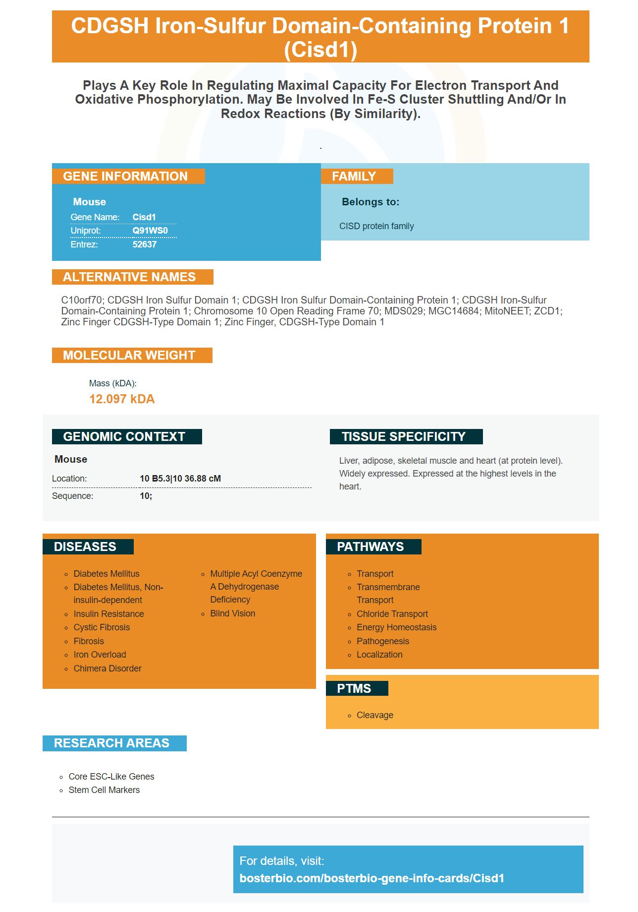
Facts about CDGSH iron-sulfur domain-containing protein 1.
.
Mouse | |
---|---|
Gene Name: | Cisd1 |
Uniprot: | Q91WS0 |
Entrez: | 52637 |
Belongs to: |
---|
CISD protein family |
C10orf70; CDGSH iron sulfur domain 1; CDGSH iron sulfur domain-containing protein 1; CDGSH iron-sulfur domain-containing protein 1; chromosome 10 open reading frame 70; MDS029; MGC14684; mitoNEET; ZCD1; zinc finger CDGSH-type domain 1; zinc finger, CDGSH-type domain 1
Mass (kDA):
12.097 kDA
Mouse | |
---|---|
Location: | 10 B5.3|10 36.88 cM |
Sequence: | 10; |
Liver, adipose, skeletal muscle and heart (at protein level). Widely expressed. Expressed at the highest levels in the heart.
When you are considering the use of the CISD1 marker in your research, you need to consider the function of the gene. There are many uses for this gene. T cell dysfunction is caused by the expression of genes called TCRs. In this article, we'll look at a few of these genes and their functions. We'll also touch on how to detect these genes.
CISD1 is an RNA interference target gene found in vertebrates. It has a C-terminus sequence that is similar to CISD2. This is an example of a protein that has an inefficient ability to bind COPI complex. Its C-terminus is devoid of a functional COPI-binding KKxx motif. Several studies have shown that this protein is an RNA interference target.
The CISD1 protein is a highly conserved gene and is expressed in all human cells. The CISD1 protein is expressed in the brain and is a component of the apoptotic caspase system. The overexpression of CISD1 has several effects, including improving neuronal viability and decreasing ROS levels. CISD1 overexpression also ameliorates the effects of HI injury in cortical neurons.
CISD1 is an ancient family of proteins that contains a 39-amino acid CDGSH domain. It is present in the outer membrane of the mitochondrion, and its activity may regulate oxidative phosphorylation and maximal electron transport capacity. It has been used as a drug target for the diabetes drug Pioglitazone. Antibodies against CISD1 may also recognize a metal molecule that binds to its protein.
The CISD1 gene expression positively correlates with the level of VAT and ATP5G3. The CISD1 marker is correlated with CD8+ T cells, whereas it negatively correlates with the level of neutrophils. The low-level CD4+ T cells and neutrophils are linked with better prognosis, while lower levels of FANCD2 predict poor prognosis.
Researchers found that CISD1 gene expression correlates with systemic insulin sensitivity. In a study of human adipose tissue, overexpression of CISD1 gene improved systemic insulin sensitivity, whereas Cisd1 knockdowns had worse glucose tolerance. This suggests that CISD1 is a good candidate for research in adipocyte differentiation. However, these findings are not the end of the story.
The complex pathway that controls the activation and differentiation of T cells is triggered by helminths, allergens, epithelial cells, and IL-4-producing innate and natural helper cells. The genes that control the functions of these cells are called effector T helper cells, or T-cells. Recent studies have characterized these cells in mice and humans.
T cells produce cytokines, a major effector of T cell mediated immunity. While most T cells lack this ability to recognize foreign substances, a subset of these cells can kill virus-infected cells. The immune system uses a combination of CD4+ and CD8+ T cells that express the so-called "Th2" cytokine profile.
LSP1 is a gene that controls T-cell migration and interaction with pERK. In a previous study, LSP1 was shown to regulate T-cell migration by deactivating pERK. The researchers found that LSP1-dependent gene repression affects T-cell function by up to 50%. These findings suggest that LSP1 is involved in T-cell function, but they don't know which gene is responsible for it.
The metabolic profile of T cells determines whether or not the T-cell response to a vaccine is effective. By targeting this gene, the immune system can better recognize and respond to the vaccine. Additionally, genetically manipulating T cells to regulate T cell function/dysfunction has many benefits. This means that we can better understand the mechanisms of vaccine-induced T cell differentiation.
The role of LSP1 in the development of T cells is unknown, but recent studies have revealed that it is required for the primary phase of infection. The homing sites of TCM cells are secondary lymphoid organs and peripheral tissues. In contrast, tissue-resident memory T cells lack migratory capacity and remain permanently in the peripheral tissues. In addition, LSP1 increases the production of autocrine IL-2, a hormone which aids the immune system in combating infectious diseases.
In addition to blocking LAG3, other immunotherapy drugs target a specific gene in the immune system known as LAG3. This inhibitor, B7S1, has shown great promise in the treatment of advanced melanoma. In fact, many LAG3-blocking drugs have been approved in the United States for the treatment of various types of cancer. This is because LAG3 is a protein that negatively regulates T cell activation. Because of its relationship to T cell activation, B7S1 inhibition suppresses tumor development in murine tumors.
The data obtained from the Boster Bio study shows that there is a complex network of gene expression that affects T cell function/dysfunction. One such gene is IRF1, which is also a target of TH9 cells. It is therefore not surprising that this gene is important in the development of a specific autoimmune disease. This reveals an important role for IRF1 in anti-tumor immunity.
In addition to its function in determining immune responses, gene expression also plays a role in regulating the number of T cells. These cells secrete antibodies to combat foreign substances. The immune response from these cells is essential in the protection of our body against infections and diseases. This response involves the activation of memory B and T cells, which survive the presence of antigens. The function of these memory cells is to produce secondary immune responses, which are more rapid than the primary response.
The gammadelta T cell/IL-17/neutrophil axis is an important new target for the treatment of cancer. However, further studies will be needed to identify how gammadelta T cell/IL-17/neutrophil axis interacts to influence immune-cell trafficking in the TME. If these findings prove to be valid, it could mean that we'll be able to design drugs that target these pathways.
In addition to these studies, the researchers have also discovered that the expression of specific homing receptors is tightly linked to the location of activation. Specifically, priming of T cells in the mesenteric lymph nodes increases the expression of the homing receptor a4b7, which facilitates migration into the gut. Similarly, the inguinal lymph nodes induce the expression of the skin homing receptors. Ultimately, this knowledge is useful in the development of vaccines.
PMID: 17376863 by Wiley S.E., et al. MitoNEET is an iron-containing outer mitochondrial membrane protein that regulates oxidative capacity.