This website uses cookies to ensure you get the best experience on our website.
- Table of Contents
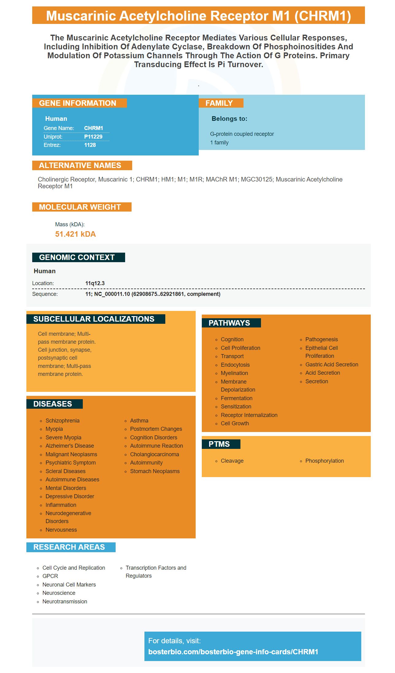
Facts about Muscarinic acetylcholine receptor M1.
.
Human | |
---|---|
Gene Name: | CHRM1 |
Uniprot: | P11229 |
Entrez: | 1128 |
Belongs to: |
---|
G-protein coupled receptor 1 family |
cholinergic receptor, muscarinic 1; CHRM1; HM1; M1; M1R; mAChR M1; MGC30125; muscarinic acetylcholine receptor M1
Mass (kDA):
51.421 kDA
Human | |
---|---|
Location: | 11q12.3 |
Sequence: | 11; NC_000011.10 (62908675..62921861, complement) |
Cell membrane; Multi-pass membrane protein. Cell junction, synapse, postsynaptic cell membrane; Multi-pass membrane protein.
The CHRM1 marker has several applications in immunohistochemistry. We will be discussing its use in IF and IHC–Fr. This is one of the most commonly used markers in immunohistochemistry. It has many other molecular biology applications. Here are some of these more common uses for the CHRM1 mark.
A good immunohistochemistry (IHC) antibody should be specific for the target protein. Non-specific staining characteristics and sensitivity are also important characteristics of an IHC antibodies. LifeSpan brand IHC antibodies should have high affinity, low stained artifacts, high signal-to noise ratio, and high staining. Furthermore, the specific staining should be stronger that the background staining.
The tissue is fixed with formaldehyde up to three hour. IHC samples require a shorter fixation process that uses less formaldehyde. This may cause epitopes to be masked and reduce antigen antibody binding. Before staining can begin, the antigen must have been extracted using a specific method. These techniques include heat-induced epitope retrieval, proteolytically-induced epitope retrieveal, and heat-induced epitope retrieveal. During sample preparation, antibodies used in IHC must first be blocked with an unspecific antigen.
In IHC-Fr, a colored compound is applied to the antigen to visualize its interaction with the target protein. Secondary antibodies are used to stain the antigen. They are labeled with a label. Several types of detection systems are common, including the avidin-biotin complex (ABC) complex, labeled streptavidin biotin, and phosphatase anti-phosphatase. The polymer based detection system has higher sensitivity.
MDAMB468-1510A cell block sections were purchased from AMS Biotechnology Europe (Massagno, Switzerland). After preparing the sections, a standard IHC protocol was applied, including deparaffinization, hydrogen peroxidase, target retrieval, and protein block. The cell block sections were processed using the traditional IHC method after scanning uIHC. After scanning uIHC cell block sections were incubated together with streptavidin oxidase (DAB substrate) and incubated for another step.
IHC-Fr requires that you follow a series of steps in order to obtain a reliable result. The sample should be properly handled and fixed. The same antigen exposure must be applied to the sample as to the validation sample or optimization sample. A competition assay using an immunizing protein must be done if the antigen level is not high. The results from multiplexed IHC-Fr should be similar, regardless of whether the antibodies are used to identify a specific antigen.
Paraffin must be completely removed for FFPE sections. Paraffinization must be completed. This will obscure the target antigens and make it difficult for antibodies to react with them. Hydrophobic properties make paraffin incompatible with aqueous solutions for IHC. Xylene is a volatile, flammable organic solvent and must be removed from the slide prior to IHC staining.
After washing the slide in PBS, it is important to perform an antigen retrieval step. The slide can be identified if the antigen is present. The protocol for antibody detection must be followed. Before IHC staining can be done, the antigen, type, and species of tissue must all be optimized. It is essential to identify the antigen as well as the antibody target. In IHC-Fr has become the gold standard in tumor analysis and is the primary technique of cancer research.
The CHRM1 marker can be used in IF to identify genes coding a receptor. This marker has been used to identify the acetylcholine M receptor. The CHRM1 gene also has several splicing variants. This marker can also help detect the presence other related genes. The development of autism is linked to the acetylcholine receptor gene.
The CHRM1 Subtype is involved with cognitive processes and can be found in brain regions linked to schizophrenia. It has been shown to be involved in these processes in knockout mice. An array of evidence has implicated dysfunctional prefrontal cortical circuitry in schizophrenia, with cholinergic deficits being specifically implicated. Because schizophrenia is closely associated with cholinergic pathways, there is no separate endophenotype.
IF-Fr is an if-else statement that can be used to test whether a value is true or false. The fr value is compatible with fixed and percentage values. When fr is used in conjunction other fixed or percentage numbers, it will be truncated to fill any space left after the other values. This makes IFFR a versatile type of data. Take the IFFR challenge to learn more about IFFR.
PMID: 3697105 by Allard W.J., et al. Sequence of the gene encoding the human M1 muscarinic acetylcholine receptor.
PMID: 2336407 by Chapman C.G., et al. Isolation of the human ml (Hml) muscarinic acetylcholine receptor gene by PCR amplification.