This website uses cookies to ensure you get the best experience on our website.
- Table of Contents
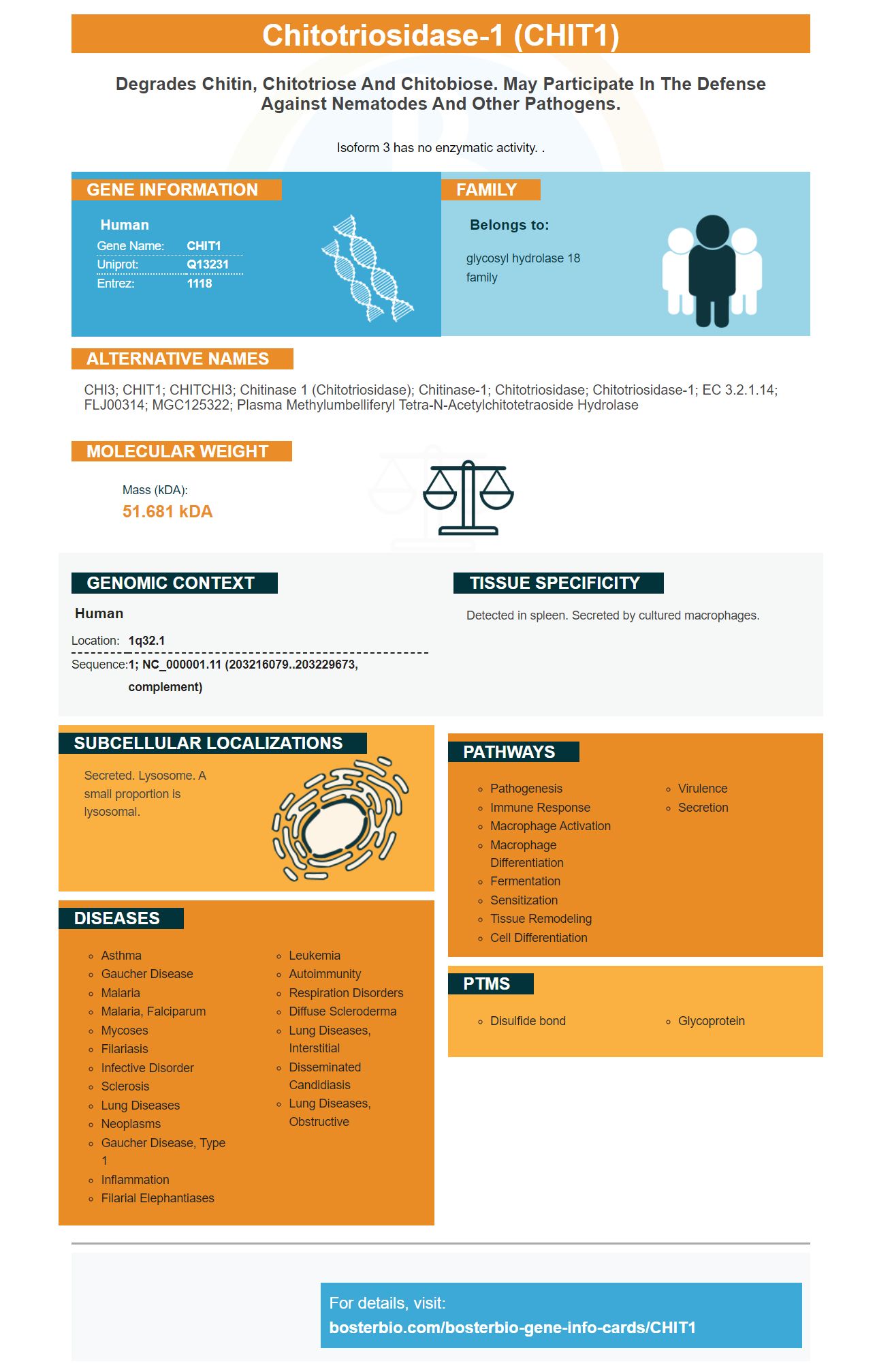
Facts about Chitotriosidase-1.
Isoform 3 has no enzymatic activity. .
Human | |
---|---|
Gene Name: | CHIT1 |
Uniprot: | Q13231 |
Entrez: | 1118 |
Belongs to: |
---|
glycosyl hydrolase 18 family |
CHI3; CHIT1; CHITCHI3; chitinase 1 (chitotriosidase); chitinase-1; Chitotriosidase; chitotriosidase-1; EC 3.2.1.14; FLJ00314; MGC125322; plasma methylumbelliferyl tetra-N-acetylchitotetraoside hydrolase
Mass (kDA):
51.681 kDA
Human | |
---|---|
Location: | 1q32.1 |
Sequence: | 1; NC_000001.11 (203216079..203229673, complement) |
Detected in spleen. Secreted by cultured macrophages.
Secreted. Lysosome. A small proportion is lysosomal.
You're not the only one who has wondered about the best uses for the CHIT1 Marker. The CHIT1 biomarker has been around for years, but only recently has it been recognized as an important biomarker for sarcoidosis. This article will discuss how CHIT1 works as a DNA damage marker and its potential utility in many applications.
The CHIT1 mark is a useful diagnostic tool for active and inactive sarcoidosis. It is highly sensitive, and it can be used to distinguish between active sarcoidosis (FUO) and active sarcoidosis (AS). Multiple studies have shown that CHIT1 levels correlate strongly with the severity and duration of sarcoidosis. This article will discuss the different uses of this marker in identifying the disease.
Patients with SMA had elevated CHIT1 levels in their CSF compared to those who were healthy. The correlation was weak and likely due to a single outlier. The CHIT1 levels did NOT vary by SMA type, SMN2 number, or patient's height. CHIT1 levels also did not differ significantly among treatment-naive patients compared to controls.
The CHIT1 marker can only be used to diagnose prion disease, AD, and frontotemporal lobar dementia. Its diagnostic value in ALS has been limited, and its levels correlated with the disease's progression. Nevertheless, CHIT1 staining was confined to a specific region of the spinal tract and was associated with markers of macrophages and microglia. However, it could not detect Creutzfeldt-Jakob disease.
CHIT1 is a biochemical indicator that can help identify lysosomal and infectious storage disorders, chronic inflammatory diseases, and RA. CHIT1 may be useful in your own research. It can also be misleading, especially for patients who do not have a thorough understanding of RA. Best Uses for the CHIT1 marker
The best uses for the CHIT1 genome damage marker are numerous and include cancer research as well as diagnostics. Its expression in human cells is higher than in non-cancerous tissues. However, statistically, these differences are not significant when compared with levels of the corresponding protein found in normal tissues. The type of sample to be tested will determine which CHIT1 DNA damage marker is most effective.
Chitotriose, which is a sugar made by some bacteria, contributes to inflammation. Chitotriose is produced by the CHIT1 gene in the human intestine and lends an advantage to some bacteria. The CHIT1 gene encodes Chitotriosidase. This compound is produced by microbes living in the human gut. Researchers used DNA sequencing for the study of the CHIT1 protein in 320 Han patients with CRC and 320 controls. Two regions, known as rs61745299 and rs35920428, were examined for their association with colorectal cancer.
Although functional data have not been collected, H. Pylori infections are believed to be responsible for the expression of CHIT1 within the gastric mucosa. The protein is involved at the hydrolysis of lacDiNAc. It is a GlcNAc–containing glycoprotein that ornaments gastric mucins. While further experimental testing is needed in order to confirm its role, the gene is still present in gastric mucins and is believed negatively to impact H. pilori adhesion.
Multiple sclerosis has been linked with activation of CHIT1. Activation of CHIT1 is associated with increased risk of multiple sclerosis and Alzheimer's disease, even though chitin is missing in humans. However, a recent study found that CHIT1 activity correlates with MS and AD. While no chitin-like substances are found in AD brains yet, it was discovered that CHIT1 activity is significantly higher in the CSF in patients with neurodegenerative diseases.
CHIT1 also plays a role as a mediator of inflammatory diseases. Recent studies have shown that CHIT1 plays a role in the metabolism chitin-like Polysaccharides across a variety tissues. Besides its role in inflammatory and infectious diseases, CHIT1 may also play a role in the production of some antigens.
There is a need to identify specific biomarkers for sarcoidosis in order to guide clinical decisions and predict clinical outcomes. Chitotriosidase could be a useful biomarker in the diagnosis of patients with this disorder. The level in CHIT1 serum was determined in a group with sarcoidosis victims with different phenotypes.
Recent studies of CHIT1 have revealed a link between the protein and COPD and ILD. These findings highlight its role in these diseases. Further research is needed to identify the precise role of CHIT1 in these diseases and to understand the mechanisms that regulate its expression and function. In the meantime, CHIT1 expression has been associated with COPD, a leading cause of death among patients with the disease.
It regulates Integrin-Linked Kinase (ILK), which is a protein that links integrins and cytoskeletal proteins. This protein has multiple roles, including regulating the phosphorylation of PI3K/Akt1/AKT1. It is involved in a downstream signaling cascade that activates mTOR. This pathway has been implicated as a cause of sarcoidosis progression and could be a therapeutic target.
Recent research identified 121 proteins which were differentially expressed between patients suffering from sarcoidosis and those who did not. The results showed that there was a difference in the expression of 121 proteins, including TGF-1, heat shock protein 90, mucin-5B, annexin, and TGF-1. TGF-b1 receptor signaling and expression in fibroblasts is important for tissue fibrosis.
Patients with MS who received escalation immunotherapy had significantly lower annual relapse rates and improved disease control. The escalation immunotherapy regimens involve high-dose corticosteroid pulses given at monthly intervals and maintenance therapy with an immunomodulatory disease-modifying agent. This treatment strategy depends on a reasonable decision-making process in order to determine the optimal level for immunomodulatory therapy.
Subjects with severe renal disease or without it build up in the brain. It is not known to cause side effects. Patients are asked to consider whether they really need this agent for every MRI. Some experts prefer to measure patients' EDSS every three month. However, there is no specific threshold for switching the therapy. The EDSS helps to evaluate a patient's reaction to immunomodulatory treatment.
The length of the disease course is dependent on your age. A relapse occurs approximately every six months. Relapse attacks must be stopped within 30 days to prevent progression to the secondary progressive forms. Between attacks, the patient can maintain a normal neurological status. Generally, pediatric MS is diagnosed at an age of 15 years. Although the disease may be more severe for adults than in children with MS, advanced diagnostic methods help to identify it earlier.
Variable immunomodulatory effects are present in the antibody response to COVID-19 injection. However, data on the individual DMTs' impact on the response to COVID-19 have been limited. The study investigators wanted DMTs examined to see if they had any effect on the antibody response. To determine this, investigators retrospectively reviewed a chart review and performed multivariable logistic regression analytics to identify factors associated for positive or negative response.
A fetal DNA test (NIPT) in the mother's blood is a non-invasive prenatal screening test that is 99% accurate at detecting a genetic abnormality in a fetus. Social Security does NOT cover this test. Women need to weigh the risk of miscarriage and the benefits of a diagnosis. Some women feel comfortable undergoing the test without further testing. If you are considering the test as a screening test for your pregnancy, your midwife, or obstetrician, will answer any questions.
A PET test is non-invasive, which means that you do not need to undergo a surgical procedure. It uses a brief-lived radioactive material. The process itself can take as long as an hour or two. The PET scanning facility generally requires patients to spend at least an hour. They will lie on a flat table that slides into a hole shaped like a doughnut in the middle PET scanning equipment.
Cell-freeDNA analysis is another type of noninvasive test. It analyzes DNA fragments that are found in the bloodstream of a pregnant lady. Most DNA is found inside the cell nucleus. Cell-freeDNA is composed of fewer 200 building blocks of DNA. They are therefore known as cell free DNA. These DNA fragments form after cells die and are released into the bloodstream.
PMID: 7592832 by Boot R.G., et al. Cloning of a cDNA encoding chitotriosidase, a human chitinase produced by macrophages.
PMID: 7836450 by Renkema G.H., et al. Purification and characterization of human chitotriosidase, a novel member of the chitinase family of proteins.