This website uses cookies to ensure you get the best experience on our website.
- Table of Contents
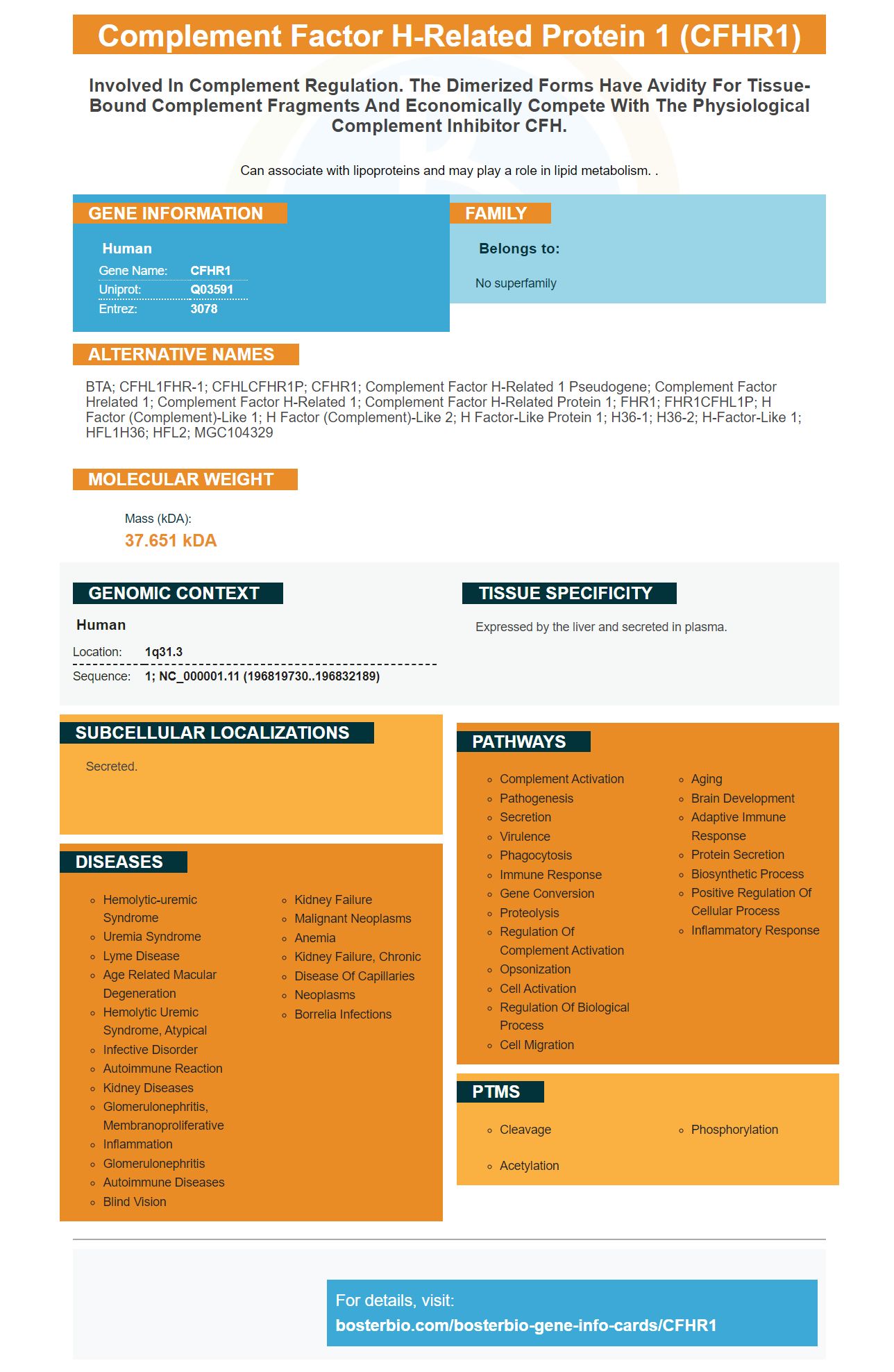
Facts about Complement factor H-related protein 1.
Can associate with lipoproteins and may play a role in lipid metabolism. .
Human | |
---|---|
Gene Name: | CFHR1 |
Uniprot: | Q03591 |
Entrez: | 3078 |
Belongs to: |
---|
No superfamily |
BTA; CFHL1FHR-1; CFHLCFHR1P; CFHR1; complement factor H-related 1 pseudogene; Complement Factor Hrelated 1; Complement Factor H-related 1; complement factor H-related protein 1; FHR1; FHR1CFHL1P; H factor (complement)-like 1; H factor (complement)-like 2; H factor-like protein 1; H36-1; H36-2; H-factor-like 1; HFL1H36; HFL2; MGC104329
Mass (kDA):
37.651 kDA
Human | |
---|---|
Location: | 1q31.3 |
Sequence: | 1; NC_000001.11 (196819730..196832189) |
Expressed by the liver and secreted in plasma.
Secreted.
Boster Bio offers flow procedure features that will optimize your experiments. There are many options available, including CFHR-1 or CFHR-E. Boster Bio's optimization tips, guides, and guides will help you decide which one is best for your experiments. Here are a few things to consider when choosing a flow procedure. This guide will describe the different flow procedures offered by Boster Bio and their respective functions.
The CFHR1 Gene is found in several tissues and organs. The CRISPR/Cas9 method was used to delete the gene from the C57BL/6 mouse. The scientists then backcrossed heterozygous mice and wild-type mice for six generations. As controls, the study used wild-type littermates aged matched to the littermates.
Cfhr1/ mice compared to wild-type controls showed that their inflammatory response was accelerated when there was no FHR-E. The plasma levels of IL-1b/TNF-a and TNF-1a were evaluated up to six hours after the LPS stimulation. Mice deficient in Cfhr1 had a higher C5 but a lower C3. FHR1/ mice also had a lower number of lymphocytes.
Despite these discoveries, the genetic defect CFHR–E has remained elusive. Genetic mutations may lead to the development of other diseases. However, a CFHR1-/ mouse model has provided an excellent tool for investigating the role of AP in inflammatory and autoimmune diseases. Patients without FHR-1 could be more susceptible to severe conditions such as sepsis.
This gene regulates AP activity. Cfhr1/ mice produce more C5a following LPS challenge. However, this effect is insufficient to influence AP regulations. Further studies are required to understand the role of FHR-E in regulating AP. In vitro studies reveal that the CFHRE gene is essential for regulating AP activation, and that it promotes the formation of C5a.
The AP pathway relies on the CFHR1 genes. FHRE is required to activate the AP pathways. FHRE is essential for the proper cleavage of C3a & C5a. Furthermore, FHR-E deficiency inhibits LPS-induced AP activation. Read on to learn more about the functions and properties of FHR–E.
This study used an anti CFHR1 antibody to measure the liver activity. Because the CFHR1 protein is highly expressed here, we used a mouse model with CFHR1 homozygous deletion. To perform the experiment, we used an RNeasy Mini Kit to obtain hepatic RNA from mice. PicoKine, a proprietary ELISA system, was used. This platform enables us to generate high-sensitivity ELISA kits.
The CFHR1 gene is expressed in the blood. It is important in sepsis. It regulates the alternative complement pathway. It has been linked to nephropathy and atypical hemolytic-uremic syndrome. The exact mechanism by AP activation is not known. This work was possible because the Cfhr1 knockout mouse was created.
Although the CFHR1 genes is found in most tissues, they are not expressed in every cell. This gene does not appear in many cells of the body, and it is not expressed in the liver of sick patients. This gene is associated with autoimmune disorders such as Lupus, which most people don't understand. This gene is essential for many processes, including immune response.
CFHR1 is a key gene in inflammatory diseases such as cardiovascular disease. It is a member of the CFH family. It has been proven that incomplete penetrance can cause the disease. CFHR1 homozygous mutation has no known cause. However, infection is necessary for disease development. Infection-related thrombotic microangiopathy is triggered by LPS, the main constituent of the gram-negative bacterial cell wall. This gene defect promotes AP activation.
CFHR-1, an essential cytokine controlling the activity T cells, is a key cytokine. Atypical hemolytic Uremic Syndrome may result from the deletion of this gene. However, IgAN and age related macular degeneration can be prevented by the deletion of this gene. CFHR-1 regulates FH and inhibits C5 converterase.
The alternative complement pathway can be controlled by the CFHR1 genetic gene. This gene is important for preventing sepsis, and other life-threatening conditions. AP activation is also controlled by the CFHR1 genes. This research was designed to identify the mechanism behind sepsis. It is likely that a new drug, which targets this gene, will be developed using these results.
FHR-E has been identified as a therapeutic target for AP activation. Its absence inhibits the activity of AP proteins, which in turn triggers the expression of C5a. Cfhr1/ mice showed elevated plasma levels of C5a after LPS stimulation. This finding was also confirmed to be an antigen in properdin.
The livers DDD-prone or AMD-prone mice have the highest concentrations CFHRC and CFHRE mRNA. In the present study, we used liver samples from cfhTg/mCfh-/-chimeric mice to determine the mRNA levels of these genes. This study also showed that CFHRE is expressed in livers of WT mice.
Previous studies have shown the competitive inhibition of CFH by the human CFHR Protein. However, the CFHR protein of mice is still not fully characterized. This study was undertaken to determine if there were differences in the levels and pathologies of complement proteins among different strains. Most complement proteins can be synthesized in liver. To assess the expression levels, liver tissues were used.
Although the function of CFHR-C is unknown, increased levels of CFHR-C mRNA in the liver of DM and DDD-prone mice suggest that the protein may be involved in the pathogenesis of these diseases. However, CFHR mRNA expression in lupus-prone mice is different suggesting that CFHR proteins play distinct regulatory roles.
Genetic analysis has revealed that there are three variants in CFHR-C, all of which encode the Fhrc genes. The specificity can be determined by excluding mouse CFHRB from qRTPCR tests. The fH/ mice were used to control, and RNA from various organs were used as templates. This approach has several advantages.
The CFHR1 molecule is found on Bruch membrane and the endothelial cells. The protein displays autofluorescence when stained using an mAb against the C5. The C terminal domain of CFHR1 appears to play a major role in binding. Surface plasmon resonance was also used for the investigation of CFHR1 binding affinities.
Immunoprecipitation experiments were used to determine the exact binding site for CFHR1 to CFHR5. To perform immunoprecipitation, human plasma was incubated with monoclonal antibodies against CFHR1. The resulting complexes were then separated by SDS/PAGE and transferred onto membranes. To confirm binding to immobilized antibodies, Western blotting was performed. In a separate experiment, CFHR1-deficient samples failed to bind to C5.
CFHR-1 in humans has been detected in three cases, all in patients with CF. The affected family had three copies of CFHR1 from probes 14 to 25. Further investigations confirmed that the affected family had three copies of CFHR1 from probes 14 to 25. A unaffected son was also a carrier. A duplication of CFHR1 was also found in a couple with two affected children, but not CFH.
C3 conversionase may be controlled by CFHR1. Incubation of activated carbon3 with factor D or factor B is required to generate C3a. This is due to the activity of C3 converterase. The experiments showed that CFHR1 at 25 mg/mL or 50 mg/mL did no significant inhibit the production of C3a. It is possible CFHR1 might inhibit C3a indirectly.
CFHR-1 belongs to the heparin protein family and is expressed on cell surfaces. It is also found in the Bruch membrane. The Cterminus of CFHR1 binds heparin C3b and cell surface. CFHR1 is likely capable of distinguishing between foreign and own surfaces. This study will concentrate on the role played by CFHR1 in the development and maintenance of the retina and choroids.
The CFHR1 & CFH protein functions as competitive antagonists for complement factor H. This regulates complement activation live. Their roles are critical in determining the mechanism for the complement system. Variation within the gene clusters can lead to disease. The study also identified CFHR1 as a predisposition for disease. The study suggests that CFHR1 may be important in regulating complement within the retina.
We don't know enough about the role of CFHR-1 and sepsis. This mouse model is a unique tool for studying AP activation. LPS-induced inflammation activates AP. The mice with FHR-E deficiency exhibit greater inflammation and reduced C5a levels. Mice with impaired FHR have AP activation in the absence of CFHR–E.
Biocytogen Co., Beijing, developed a gene editing method called CRISPR/Cas9 to delete CFHR-1 from mice. Mice were bred in a specific pathogen-free facility. This technique allows the team determine if the deletion has been confirmed to be stable and functional at a genomic level. If confirmed, the research will allow for a more accurate diagnosis of CFS/CFHR-1.
PMID: 1826708 by Estaller C., et al. Cloning of the 1.4-kb mRNA species of human complement factor H reveals a novel member of the short consensus repeat family related to the carboxy terminal of the classical 150-kDa molecule.
PMID: 10781834 by Male D.A., et al. Complement factor H: sequence analysis of 221 kb of human genomic DNA containing the entire fH, fHR-1 and fHR-3 genes.