This website uses cookies to ensure you get the best experience on our website.
- Table of Contents
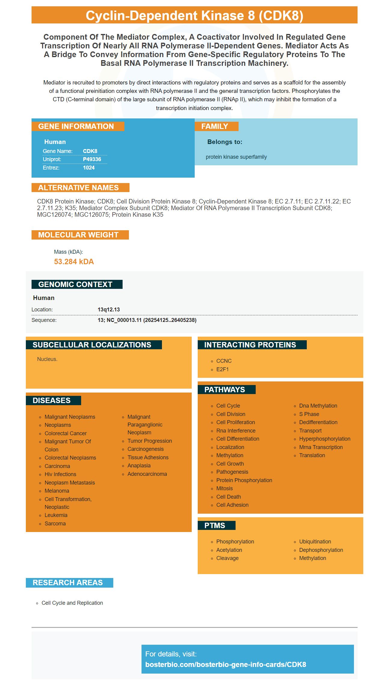
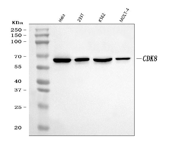
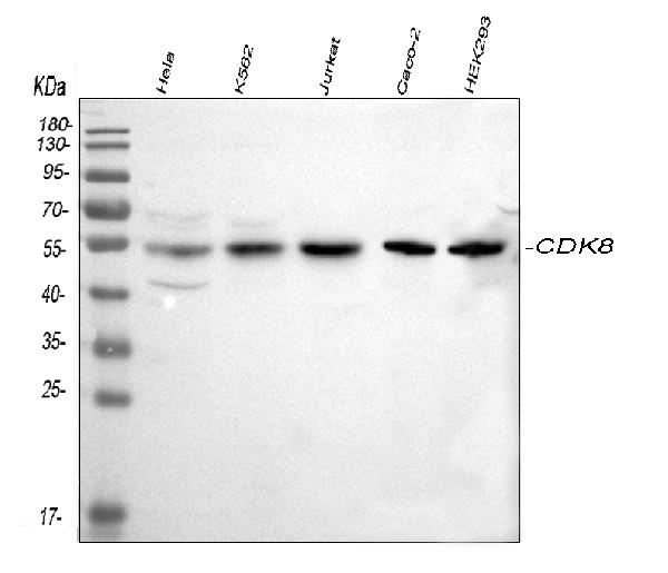
Facts about Cyclin-dependent kinase 8.
Mediator is recruited to promoters by direct interactions with regulatory proteins and serves as a scaffold for the assembly of a functional preinitiation complex with RNA polymerase II and the general transcription factors. Phosphorylates the CTD (C-terminal domain) of the large subunit of RNA polymerase II (RNAp II), which may inhibit the formation of a transcription initiation complex.
Human | |
---|---|
Gene Name: | CDK8 |
Uniprot: | P49336 |
Entrez: | 1024 |
Belongs to: |
---|
protein kinase superfamily |
CDK8 protein kinase; CDK8; Cell division protein kinase 8; cyclin-dependent kinase 8; EC 2.7.11; EC 2.7.11.22; EC 2.7.11.23; K35; Mediator complex subunit CDK8; Mediator of RNA polymerase II transcription subunit CDK8; MGC126074; MGC126075; Protein kinase K35
Mass (kDA):
53.284 kDA
Human | |
---|---|
Location: | 13q12.13 |
Sequence: | 13; NC_000013.11 (26254125..26405238) |
Nucleus.
This article will focus on Boster Bio and its many uses. We'll also go over its validation and quality of the Reagent Container. We'll also discuss the advantages and disadvantages of the CDK8 marker. You'll be able find the perfect antigen for your research in just an hour. Learn more about Boster Bio and what you can expect.
The Cdk8 gene encodes the protein that phosphorylates serines within the PASdomain. PAS domains detect changes in the environment and convert them into the appropriate cellular response. They use the heme hormone in mammals to detect O2. PAS domains are characterized by an amino acid sequence with a tiny homology. The three-dimensional fold of PAS is highly conserved. It consists of a five-stranded antiparallel sheet, and two a-helices.
The CDK8 gene can be used to determine the genetic status of cancer cells. The CDK8 gene is found in a variety of tissues including colon cancer. The TCGA database includes cancer cell lines. In a few experiments, cells were treated with interferon type II grams at 2.5 ng/ml. Below are some examples of the CDK8 marker. In-depth data collection and analysis methods are described in the accompanying materials and methods.
SEL120-34Ais a novel CDK8 inhibitor, is characterized by its potency and selectiveness. It binds to CycC and forms a double-stranded helix with the CDK8 pocket. It also inhibits STAT1 S726 phosphorylation. The SEL120-34A inhibitor also blocks the phosphorylation process of RNAPII CTD S2 as well as MCL1, two additional transcription factors in leukemia cells.
CDK8 inhibitors can also be used to reduce the activity of tumor-promoting paracrine cells. This inhibitor decreases the paracrine functions that promote tumors in normal fibroblasts. Treatment of cancer cells caused by DNA damage CDK8 inhibits has been proven to improve the chance of survival. In the absence of an CDK8 inhibitor, this treatment might not be effective. This is due to the fact that CDK8 inhibition has been demonstrated to correlate with poor survival rates and has been associated with a poor prognosis for breast and ovarian cancers.
The deletion of the CDK8 gene provides many other benefits. Cells lacking the enzyme exhibit a distinct appearance and phenotype than those with the SSN801 mutation. In addition, cells which do not contain Cdk8 display a morphological change in mitochondria and a lower virulence. Additionally, Cdk8 deletion reduces the cell's virulence in the same compartment, indicating that the two genes function independently of one another.
CDK8 is a member of the kinase family. Its T-loopthat is vital for kinase activity and stable interaction with substrates, is crucial. Pathogenic mutations can alter the T-loop's structure dynamics or its binding to substrates. The CDK8 gene is encoded by two genes, Cdk8 and MED12. The gene product encodes a protein vital for the kinase activity of this kinase.
Congenitively cardiac overexpressions of CDK8 can cause premature lethality in mice. Cdk8 overexpression causes an increase in the expression of genes involved in oxidative metabolism and an increase in the cytoskeletal forms of cardiomyocytes. In addition, overexpression of CDK8 triggers cardiac fibrosis. Therefore, the confirmation of the CDK8 marker requires further research. The cellular distribution of CDK8 has been investigated using QRT-PCR.
The CDK8 gene is a key part of the Mediator transcription complex. It is a major target of drugs as it plays a role in the regulation and expression of a variety of types of genes. CDK8 is part of a large CKM complex and requires the presence of CKM to function as an Kinase. Its overexpression promotes the induction of silent genes and the downregulation of mitochondrial-related pathways.
The protracted survival curve of mice infected by CDK8/CDK9 double-knockouts was more extensive than that of their WT counterparts. They were also less capable of crossing the blood-brain barrier. The results of these experiments were representative of two independent studies. The CDK8/CDK19 IC50 levels are listed as mean + standard deviation. To confirm the CDK8/CDK19 inhibitors, the IC50 values must be determined.
The Boster Bio Reagent Quantity Container includes the following items: 96 Well Cell Culture Clear-Bottom Microplates, 10x TBS, 1x Clear Quenching Buffer and 1x Blocking Buffer. This product also contains 100x Rabbit Polyclonal Antibody. For optimum performance, make use of this antibody with the 96-Well Cell Clear-Bottom Microplate.
When receiving the product take the time to inspect the package and the contents. Contact Boster Biological Technology immediately if you discover any damage, shortage or any other defect. Note that specific shipping instructions may apply to customized products. Returns of custom products should be made in the original packaging. When you receive the product, keep it in a cool place at 4 degrees Celsius. Do not make use of the product for more than two weeks. It is recommended to throw away any product that is affected by.
If you purchase Products from Boster Biological Technology, please take into consideration that these reagents will be sold and distributed by third parties. These products are intended to be used for research, diagnostic or therapeutic use. While they might be beneficial but you should not apply them to therapeutic uses. They can trigger allergic reactions or harm to humans. Before using this reagent in your laboratory ensure that you consult your physician.
Boster Biological Technology will replace or refund the purchase if it does not meet the requirements. In these instances you should contact Boster Biological Technology and provide details of the problem. Boster will review your plan, offer reasonable advice and, if necessary, replace it. The process will take minimum two weeks however, it could take longer.
Live cell imaging is possible using SiR-based probes. They are highly permeabilized, which allows them to enter difficult to transfect cell types such as erythrocytes. Further, they are fluorogenic that allow for direct detection of probes that are bound to targets without the need for washing steps. This makes them able to be used for a variety of applications, from tissue imaging to cell biology.
Scientists were able use SiR-derivatives to link synthetic fluorescent probes to connect proteins. The use of SNAP-based Fusion proteins in live cells, for example could not have been possible without far-red fluorophores. Researchers developed SiR-SNAP, the fluorescent protein that enabled researchers to label SNAP-expressing neurons within sections of the brains of rats.
Live cell imaging is a popular application for the staining of cells permeable to DNA. The current DNA stains have one major disadvantage. These stains are toxic and require blue light illumination. They also don't work well with super-resolution microscopy. SiR-Hoechst is a better alternative. It is a far-red DNA stain. In addition , it is highly compatible with super-resolution microscopy, it is also relatively safe. SiR-Hoechst is highly compatible different types of cells and can be used to live cell imaging.
Live cell imaging requires a non-toxic, biocompatible , and fluorogenic fluorescent probe. It should have emission and excitation wavelengths that are in the far-red. It must be able to identify biomolecules that have exceptional specificity without the need for genetic manipulation. However, only a few of these probes have been reported in the literature. This is because far-red synthetic fluorophores exhibit poor membrane permeability and are unspecific for cellular components.
PMID: 7568034 by Tassan J.-P., et al. Identification of human cyclin-dependent kinase 8, a putative protein kinase partner for cyclin C.
PMID: 9734358 by Sun X., et al. NAT, a human complex containing Srb polypeptides that functions as a negative regulator of activated transcription.