This website uses cookies to ensure you get the best experience on our website.
- Table of Contents
1 Citations 3 Q&As
Facts about CD63 antigen.
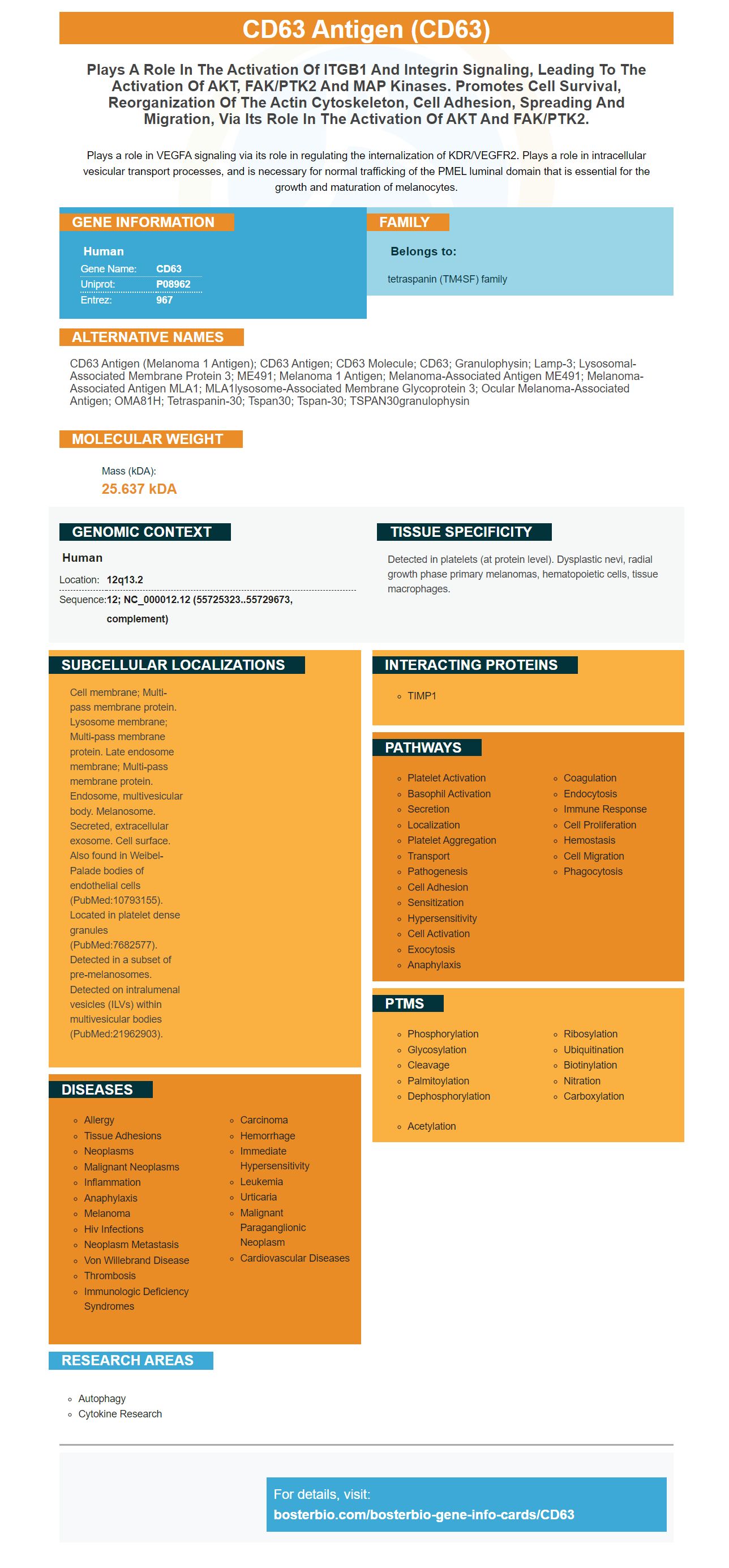
Plays a role in VEGFA signaling via its role in regulating the internalization of KDR/VEGFR2. Plays a role in intracellular vesicular transport processes, and is necessary for normal trafficking of the PMEL luminal domain that is essential for the growth and maturation of melanocytes.
Human | |
---|---|
Gene Name: | CD63 |
Uniprot: | P08962 |
Entrez: | 967 |
Belongs to: |
---|
tetraspanin (TM4SF) family |
CD63 antigen (melanoma 1 antigen); CD63 antigen; CD63 molecule; CD63; Granulophysin; Lamp-3; Lysosomal-associated membrane protein 3; ME491; melanoma 1 antigen; Melanoma-associated antigen ME491; melanoma-associated antigen MLA1; MLA1lysosome-associated membrane glycoprotein 3; Ocular melanoma-associated antigen; OMA81H; tetraspanin-30; Tspan30; tspan-30; TSPAN30granulophysin
Mass (kDA):
25.637 kDA
Human | |
---|---|
Location: | 12q13.2 |
Sequence: | 12; NC_000012.12 (55725323..55729673, complement) |
Detected in platelets (at protein level). Dysplastic nevi, radial growth phase primary melanomas, hematopoietic cells, tissue macrophages.
Cell membrane; Multi-pass membrane protein. Lysosome membrane; Multi-pass membrane protein. Late endosome membrane; Multi-pass membrane protein. Endosome, multivesicular body. Melanosome. Secreted, extracellular exosome. Cell surface. Also found in Weibel-Palade bodies of endothelial cells (PubMed:10793155). Located in platelet dense granules (PubMed:7682577). Detected in a subset of pre-melanosomes. Detected on intralumenal vesicles (ILVs) within multivesicular bodies (PubMed:21962903).
A ELISA kit is an excellent choice if you're looking to use the CD63 Marker in your research. A CD63 ELISA Kit has many advantages. The kit can be utilized in all kinds of experiments. It can also be customized to fit the needs of your research. This kit doesn't require minimum order quantities. It is applicable to researchers from all countries. For more details, visit Boster Bio's website.
The identification of tumor-associated antibodies is possible using aptamers, which were designed with the CD63 protein. A variety of advantages of aptamers over antibodies have been mentioned in the design of aptamers. For instance, aptamers can be used to identify extracellular vesicles associated with tumors from cells in a mild environment. Aptamers also target small molecules, sugars, and lipids. Furthermore, aptamers can be enhanced by the use of artificial or modified nucleic acid, as well as counter-SELEX.
Extracellular vesicles, components found in mammalian fluids have been the focus of research as biomarkers for various kinds of diseases. By using next-generation sequencing techniques, RNA aptamers were identified. Two conserved motifs were used to determine the sequences. Most predicted a nonconserved loop structure or stem structure. CD spectroscopy confirmed the binding affinity of the Aptamers.
By using the anti-CD63 aptamer of ExoPCD's microfluidic chip Xu and colleagues. (145) were able to identify CD63 proteins on exosomes using a LOD of 4.39 x 3 particles/mL. The aptamer was also suited for the detection of exosomes using immobilization-free and label-free EC system.
DNA sequences with a primary amine group at the 3' end were purchased from Integrated DNA Technologies. They were dissolved in water to a concentration of 10 mM. They contained sequences corresponding to CD63, CD24, and EpCAM. The aptamer sequences were labeled with three fluorescent molecules and then added to a four-arm poly(ethylene glycol) with free amines.
The authors also attempted to shorten the full sequence of the CD63 protein. In addition, they chose sequences that contained nonconserved stem regions. These sequences had higher peaks in the respective rankings than the remaining candidate aptamers. In addition, these sequences had longer stems and were more stable. Thus, they were selected as representative aptamers. In this way, they could be used to target the CD63 protein.
The process of selecting high-affinity aptamers has been developed in three laboratories. The SELEX method, whose name stands for systematic evolution of ligands by experiential enrichment, is used. This method yields artificial nucleic acids called aptamers. They are useful for the identification of various clinically relevant biomarkers. Aptamers can detect different targets, including proteins, amino acids, and thin slices of tumors.
The CD63 Marker is used to monitor the activity and response of the disease. It is an invaluable diagnostic tool for the diagnosis of CSU. The cost of this test varies widely, depending on the laboratory used and the number of patients tested. The use of this marker can improve the management of CSU in patients who have been diagnosed with this disorder. The CD63 Marker is a noninvasive blood test that can be used to diagnose CSU in children.
It has been shown to interact with several proteins and cell processes. It has been found to interact with a protein known as tissue inhibitor of metalloproteinase-1. Originally, this protein inhibited cancer growth but later proved to have cancer-promoting properties. In addition, this protein is involved in the activation of basophil granulocytes. Several other diseases and disorders are associated with CD63, including hermansky-Pudlak syndrome and lysosomal storage pool deficiency.
The cost of CD63 Marker depends on the laboratory used to test your samples. The more expensive the test, the more expensive the procedure. The more accurate it is, the better your treatment. The test also provides you with a report on how well the cancer is progressing. For more information, visit the CD63 marker page on our website. Our goal is to help you find the best price for your CD63 Marker. When choosing a lab, always make sure you have all the information you need, including your test results.
One of the most important tests for CSU is CD63. It shows the greatest correlation between CD63 and basophil degranulation. In addition, CD63 expression on basophils correlates with ASST in autoimmune CSU, urticaria, and response to therapy in atopic diseases. The price of CD63 Marker depends on the lab's cost-of-analysis. The test can cost as little as $25.
A new screening method for aptamers has been developed to screen for the presence of molecular markers in cancer cells. This method uses a cell-SELEX screening platform, which is unique in its ability to generate a panel of aptamers while not requiring any prior knowledge of a cell's molecular signature. Cell-SELEX-screened aptamers have high affinity and specificity for cancer cells, which may enable broader applications of the technology in disease treatment. Current applications of cell-SELEX-screened aptamers include liver, lung, and brain cancers.
A new chemical antibody, called an aptamer, is being developed, which has several benefits over antibodies. The aptamer is smaller, more readily synthesized, and more easily manipulated than antibodies. This versatility and ease of synthesis has made it a potential target-designed cancer drug. Scientists have also designed new aptamer-drug conjugates, including phototherapy and chemotherapy.
Using DNA-silver nanoclusters, Yin et al. developed a label-free aptasensor that uses fluorescent DNA-silver nanoclusters as recognition probes. This aptamer is designed to recognize cancer cells by binding to the target's DNA. The aptamer's fluorescent property is changed in the presence of cancer cells, and the corresponding chemotherapeutic effect is carried out.
Despite being an excellent drug delivery system, many chemotherapeutic agents have toxicity and poor selectivity when applied to tumor cells. By using an aptamer-drug conjugate, researchers could directly target cancer cells with a single, small dose. They would also be able to avoid toxicity to healthy cells in the body, reducing the dose and enhancing therapeutic efficacy.
The aptamer-nanoparticle preparation method can extend the half-life of the drug in blood. Nanoparticles that have a high specific surface area and uniform morphology could improve drug delivery and distribution. In recent years, nanomaterials comprised of various materials, such as silica nanoparticles made of gold, silica, and aptamer-targeted drug models, have been developed.
A novel aptamer-nucleolin molecule called AS1411 was discovered by scientists in Korea and is currently being tested in clinical trials for leukemia. It inhibits the nuclear factor-kB function and destabilizes BCL-2's mRNA. In addition, aptamers exhibit high specificity for cancer and other tumors and are extremely resistant to different types of nuclease deficient medications.
The ELISA assay for CD63 markers is a simple and affordable method to determine the amount of human CD63 in serum, plasma, or tissue homogenates. The sensitivity and specificity of the CD63 assay are exceptional, and there isn't any significant cross-reactivity between human CD63 and any of its analogs. Biotin-antibody (1x) is added to the wells.
The ExoELISA Ultra CD63 kit significantly increases the sensitivity of detection of exosomes. It can also cut down the time of the assay by only four hours. The ExoELISA-ULTRA CD63 kit is compatible with almost all biofluids and comes with a calibrated internal standard for exosome detection. This kit includes all the necessary reagents, as well as the assay plate.
A commercial ELISA assay for CD63 Markers has multiple benefits. It can detect high levels of this protein in a variety of samples, from whole blood to human tissues. It also helps in screening patients for multiple cancers by identifying their cancer-related risk factors. Cost of ELISA assay for CD63 Marker increases when more than one sample is tested at the same time.
PMID: 3365686 by Hotta H., et al. Molecular cloning and characterization of an antigen associated with early stages of melanoma tumor progression.
PMID: 2171551 by Rapp G., et al. Characterization of three abundant mRNAs from human ovarian granulosa cells.
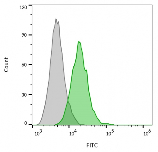
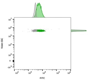
CD63 Antibody is a monoclonal antibody that has been produced against CD63. It is used for isotype control. The primary and secondary antibodies are mouse IgG and phycoerythrin-conjugated. The antibody was made from PBMCs from human. The PBMCs were fixed in paraformaldehyde before being permeabilized with saponin. Then the cells were incubated in the dispersed anti-human CD63 Monoclonal antibody.
The CD63 antibody recognizes the protein that is on the cell's surface. It is expressed on the membrane of the exosome and is activated in the normal course of cell development. It is also expressed on the cell surface by monocytes and platelets. In humans, it is present on the surface of granulocytes and macrophages, and is weakly expressed in granulocytes. The antigen is the same as the antigen that causes melanoma ME491 and the platelet antigen PTLGP40. Abcam has been a leader in reproducibility and has knockout cell lines as well as gold standard validation.
There are some caveats to using an anti-CD63 antibody in clinical settings, the most significant is patient compliance. The CD63 BAT can be used to identify the presence of autoimmune CSU patients. While it is not as sensitive as an IgG however, it is much more sensitive and specific than monoclonal antibodies which are ineffective in patients suffering from inflammatory intestinal disease or Psoriasis.
The CD63 antibody, which is a tetraspanin, is a member of the LAMP-3 superfamily. It is extensively expressed in the extracellular space , and is found on the plasma membrane following activation. It is important to be aware that interactions between CD63-adaptor proteins regulate cell activity. The 67% amino acid sequence identity of the human CD63 anti-rat CD63 and mouse CD63 is shared by both species. Although the functional profiles of the two species are identical but their clinical significance is unknown.
The CD63 Antibody monitors the disease's activity and response. It gives information about the CSU and the treatment it offers. Different studies have proven that the CD63 antibody is a powerful method of monitoring the activity of the disease. This way the CD63 Antibody can provide useful information. The immune system is largely affected by the level in the blood of CD63. CD63 is extremely sensitive to antibodies. It is essential to stay clear of negative side effects from this medication.
The CD63 antibody, a non-specific anti-inflammatory antibodythat detects the presence of apoptotic cells inside the body of a cell. It is an important part of the immune system. It is essential to maintain the health of the cell. It could indicate the presence or absence of apoptotic cells. In addition, it could aid in determining the severity of inflammation. The antibody can also boost the immune system's ability to fight.
The CD63 level is associated with the Atopy. The high level of CD63 means higher disease activity. In a study involving pediatric patients and their parents, the CD63 BAT levels were lower than those of non-atopic patients. The number of lymphocytes found in the lung is linked to CD63 levels. They were also related to disease activity based on the UAS7 score. The high antibody level suggests that aphluorin was present in the immune system.
The aim of the study was to establish a reference range for the CD63 BAT results that are based on expression for children with no CSU. The aim of the study was to evaluate the impact of gender, age, ethnicity and disease activity on the BAT results. The researchers concluded that the high expression levels of the patient population were significantly different from those of the controls. This is statistically significant but the Wilcoxon rank sum difference was less than 0.23 for controls and patients with PU.
In a recent study 23 patients in the pediatric population with PU were tested for the expression of CD63. The majority of the patients were males and had an IgE of 947 mg/L. The median CD63 levels in patients was 1.88 percent and is similar to the median value in a study with healthy subjects. In comparison to the controls, the BAT values did differ statistically between PU patients and the controls. The Wilcoxon rank sum difference was 840 and p = 0.23 respectively.
25682-1-AP, a human CD63 antibody was utilized to test the CD63 protein in CSU patients. To determine the amount of CD63 expression in human patients the anti-CD63 antibody (25682-1-AP) was employed. The dilution of the anti-CD63 was 1:800, and the samples were examined using an 80x lens, and incubated overnight at 4 degrees Celsius. The CD63 dilution in the control cells was similar to the value of the reference samples.
*More publications can be found for each product on its corresponding product page