This website uses cookies to ensure you get the best experience on our website.
- Table of Contents
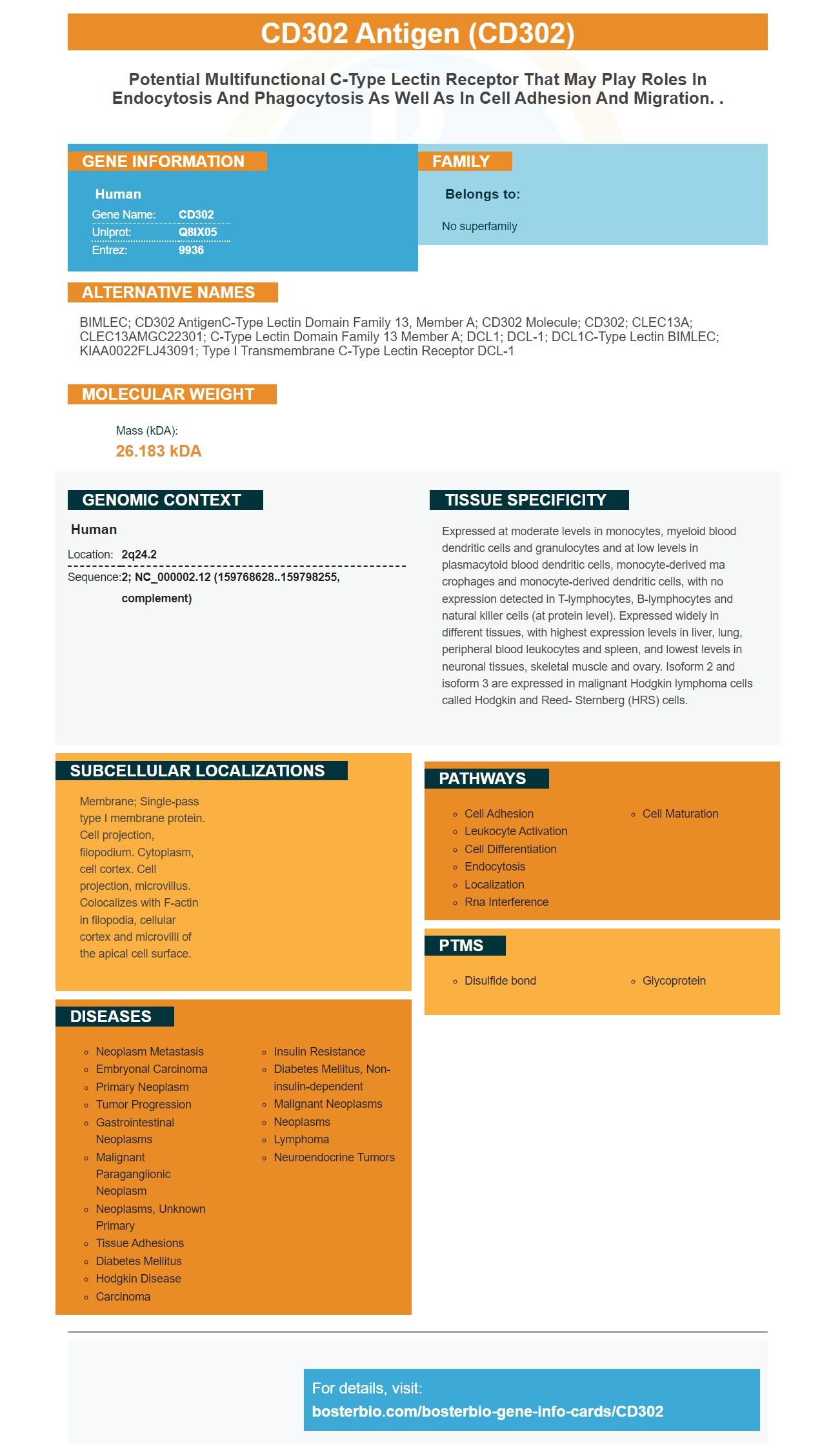
Facts about CD302 antigen.
Human | |
---|---|
Gene Name: | CD302 |
Uniprot: | Q8IX05 |
Entrez: | 9936 |
Belongs to: |
---|
No superfamily |
BIMLEC; CD302 antigenC-type lectin domain family 13, member A; CD302 molecule; CD302; CLEC13A; CLEC13AMGC22301; C-type lectin domain family 13 member A; DCL1; DCL-1; DCL1C-type lectin BIMLEC; KIAA0022FLJ43091; Type I transmembrane C-type lectin receptor DCL-1
Mass (kDA):
26.183 kDA
Human | |
---|---|
Location: | 2q24.2 |
Sequence: | 2; NC_000002.12 (159768628..159798255, complement) |
Expressed at moderate levels in monocytes, myeloid blood dendritic cells and granulocytes and at low levels in plasmacytoid blood dendritic cells, monocyte-derived ma crophages and monocyte-derived dendritic cells, with no expression detected in T-lymphocytes, B-lymphocytes and natural killer cells (at protein level). Expressed widely in different tissues, with highest expression levels in liver, lung, peripheral blood leukocytes and spleen, and lowest levels in neuronal tissues, skeletal muscle and ovary. Isoform 2 and isoform 3 are expressed in malignant Hodgkin lymphoma cells called Hodgkin and Reed- Sternberg (HRS) cells.
Membrane; Single-pass type I membrane protein. Cell projection, filopodium. Cytoplasm, cell cortex. Cell projection, microvillus. Colocalizes with F-actin in filopodia, cellular cortex and microvilli of the apical cell surface.
Optimizing flow procedures can be difficult. A guide or optimization tip can help. We will be discussing CD302 mAb expression on leukemic blood monocytes and CD302 expression upon leukemic blasts. We'll also discuss PBD delivery using CD302 Abs and some concerns about hepatotoxicity.
CD302 is a cytotoxic protein expressed on the surface of leukemic cells. It has a restricted expression profile in leukemic and myeloid AML cells. This could be due to its cellular interaction with the CD33 genes. In a study of 460 AML patients, CD302 expression was correlated with that of CD33. Most of the CD302-positive cells were found on the M4-M5 subtype, which showed the highest expression level.
To study CD302 expression during leukemic blasts, the researchers used GSE14468, a microarray dataset from Gene Expression Omnibus. The dataset includes primers to human CD302 qPCR and HPRT qPCR. The study resulted in the expression of the CLL-1 gene in leukemic blood cells from five of six patients (46%) Interestingly, one patient with refractory anemia had a negative CLL-1 expression, but two of three patients with excess blasts-2 were positive. The researchers also looked at patients with myeloproliferative diseases (MDS) or therapy-related myeloproliferative diseases.
CD302 can be expressed on leukemic blasts that have been exposed to cytotoxic cells. The fusion gene is more effective that DT388IL-3. It isn't yet clear if this protein is effective in treating the leukemia. If it is, it will be useful for monitoring treatment. It may be an important biomarker for detecting leukemia or other blood disorders.
Another study focused on the role of CD302 on hematopoietic stem cells. Taussig and colleagues isolated CD34+/CD38 stem cells from bone and cord blood. These cells are rich in HSCs, which can be used to repopulate SCID mice for long-term. CD123 expression was also found to be lower than in CD34+/CD38-cells.
Other chemokines may also promote CD302 in leukemic cellular cells. IL-3Ra expression is often high in leukemic blasts. However, IL-3Ra excess does not correlate with a better prognosis.
Recent studies have found that cord blood monocytes show differential gene expression. Although myocardial injury or sudden death is the most common cause of coronary heart disease, more long-term studies are needed in order to determine whether CD11c is associated with more severe forms of atherosclerotic disease. If the data proves to be accurate, we may expect to see a direct correlation between monocyte CD11c expression and the development of coronary artery disease.
Although the term "intermediate Monocyte" is still not defined, it has been the subject to a lot of research since 2010. A search on Google Scholar reveals that there are now more than one hundred such studies on the topic. Numerous studies have shown that these cells can predict the outcome of various diseases, including heart disease. Other studies suggest that CCR2 and other markers might be useful in identifying intermediate monocytes.
To test monocyte surface AG expression, a flowcytometer is used. Flow cytometry was used for measuring the percentage of CD14+monocytes. Positive expression was defined as a percentage of CD14+monocytes greater than 90%. CD14 Abs were used to provide negative controls. Each donor was given one sample of blood with each type of blood. The absolute number was 0.32x10⁹ cell/liter in both wholeblood and mononuclear blood samples.
HLADR expression on monocytes was studied in a study involving women. The two groups were pre and postmenopausal women. The results showed significantly higher expression levels of HLADR in postmenopausal females with cervical cancer than in premenopausal ladies. The difference was borderline significant. This study is still in progress. The authors wish to thank the volunteers for their participation in the study.
Other studies have examined the effect of CD54 on peripheral blood monoocytes in cancer. These findings suggest that CD54 is more prevalent in the blood of cancer patients. It also showed that HLA-DR was less prevalent in patients with cancer than it is in non-cancer patients. These findings have implications on the clinical diagnosis of cervical carcinoma. The study concludes that CD54 expression plays a critical role in the immune response to the tumor microenvironment.
Evaluation of PBD delivery via CD302 masb in leukemic and hepatic cells is done. It has been demonstrated that the antigen can cause cell suicide when it is paired with a secondary immunoglobulin. Furthermore, PBD can induce apoptosis and promote tumor regression when delivered through CD302 mAb. We can successfully kill cancer cells using antigens that are against CD302 in PBD delivery.
The CD302 antibody was found to be high in leukemic blood cells. These cells were all CD367+ and CD371+. Monocytes, however, expressed a high number of CLRs, including CD302, which corresponds to activating and inhibitory CLRs. The CD302 mAb was also negative in monocytes but highly expressed in cord blood and BM.
MMRI-20 was used as a detection tool to detect PBD delivery via CD302 Mabs. MMRI-20 inhibited the killing of cells in HepG2 or HL-60 cells in vitro. It was then compared to isotype control mAb and untreated control cells. We also tested whether PBMC could be killed in co-culture with 1nM GAM IgG mAb or 5% BSA TBST.
This antibody is also highly expressed on AML cells and the enriched CD34+ population. Although the CD302 anti-CD302 antibody is a potential therapeutic target in AML (although further research is required to determine an optimal therapeutic window). These results also show the utility of CD302 mAb for patients with leukaemia.
AML can be studied in vivo by targeting CD302 with an antibody. It can recruit immune effector cells and inhibit critical functions of AML. The mAb MMRI-20 is designed to target CD302 in leukemia cell lines and reduce their engraftment. The high levels of natural killing activity in the naked mouse antibody could have limited the ADCC functionality of CD302 mAb. That would explain why it failed to eliminate HL-60 cells from the host cells.
CD302 is a potential therapeutic target for AML. However hepatotoxicity concerns are still a concern for CD302 Abs. While this protein is present on the surface hepatocytes and sinusoidal epithelial cells, its MW in liver cells differs from myeloid cells. In contrast, Western blot analyses of the leukemic cell line HL-60 showed abundant CD302 expression on the surface, while HepG2 cells expressed only trace amounts of the protein.
In studies, CD302 mAbs were used to inhibit the migration of leukemic and hepatic cells. Although the mechanism of action is not known, it has been shown the anti-AML antibody mAbs recruit immune effector cell and inhibit AML cells' crucial functions. MMRI-20 binding didn't alter the migration of leukemic blood cells in vitro, but it inhibited it in other experimental situations. Future studies will identify CD302's binding ligand and explore the anti AML mAbs' blocking actions.
PMID: 12824192 by Kato M., et al. Hodgkin's lymphoma cell lines express a fusion protein encoded by intergenically spliced mRNA for the multilectin receptor DEC-205 (CD205) and a novel C-type lectin receptor DCL-1.
PMID: 17947679 by Kato M., et al. The novel endocytic and phagocytic C-Type lectin receptor DCL-1/CD302 on macrophages is colocalized with F-actin, suggesting a role in cell adhesion and migration.