This website uses cookies to ensure you get the best experience on our website.
- Table of Contents
16 Q&As
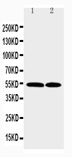
Facts about C-C chemokine receptor type 3.
Receptor for a C-C type chemokine.
Binds to eotaxin, eotaxin-3, MCP-3, MCP-4, RANTES and MIP-1 delta.Then transduces a signal by increasing the intracellular calcium ions level. Alternative coreceptor with CD4 for HIV-1 infection.
Human | |
---|---|
Gene Name: | CCR3 |
Uniprot: | P51677 |
Entrez: | 1232 |
Belongs to: |
---|
G-protein coupled receptor 1 family |
b-chemokine receptor; CC chemokine receptor 3; C-C chemokine receptor type 3; C-C CKR-3; CCR3; CCR-3; CD193 antigen; CD193; chemokine (C-C motif) receptor 3; CKR3CC-CKR-3CMKBR3CD193; eosinophil CC chemokine receptor 3; Eosinophil eotaxin receptor; MGC102841
Mass (kDA):
41.044 kDA
Human | |
---|---|
Location: | 3p21.31 |
Sequence: | 3; NC_000003.12 (46210699..46266706) |
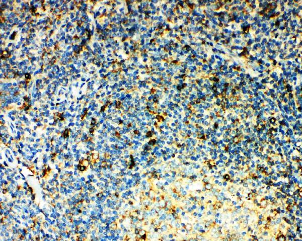
In eosinophils as well as trace amounts in neutrophils and monocytes.
Cell membrane; Multi-pass membrane protein.
In the midst of the numerous antibodies available on the market CCR3 is a notable candidate for the monitoring of sepsis. Read on to find out more about CCR3's clinical use and its biological significance in sepsis. Here are some helpful suggestions to use this marker in a clinical setting. Learn how to make your own antibody that can detect this cellular marker. This is a brief guide to help you get started.
There are a variety of biomarkers to be found that can indicate whether a patient suffers from sepsis. CCR3 is one of these potential candidates. The study was published in the Br Med J. In the study, Moore JJ, McKinley BA, and Turner K examined the epidemiology of sepsis in general surgical patients. They found that patients with CCR3 deficiencies were more susceptible to sepsis than patients with other types.
Septic shock is caused by the CCR3 molecule. In this case, the immune response leads to tissue damage and the body's T cells shift to the Th2 profile. Eotaxin is an additional ligand for CCR3. Both proteins are key chemokines in Th2/allergic processes. CCR3 and Cytokine can be activated and then migrate into areas of inflammation are Th2 cells that stimulate the humoral immune response.
Despite its distinctiveness, PCT's many clinical applications are largely unexplored. One of the problems with PCT measurement is that the levels are not yet sufficient enough to determine the severity and extent of bacterial infections. The level of PCT might not be enough to differentiate between microbial and nonmicrobial infections. A combination of biomarkers might prove more useful in clinical applications. It may be difficult to create biomarker with a reliable technology.
CCR3 is a brand new blood test that can be used to monitor sepsis. It can be used alone or with PCT, and is more reliable than CRP. It is a way to check the health of individuals of all different ages. In the end, this test can lower the amount of antibiotics prescribed, duration, and the costs associated with antibiotics. Because of its high sensitivity, it could become an important element of a sepsis surveillance program.
The severity of sepsis will be contingent on the severity of the infection. Severe sepsis usually comes with hypoperfusion or dysfunction in at least one of the organs. In severe cases, organs can be severely compromised. This condition is also referred to as septic shock. It is responsible for more than 60% of deaths in hospitals. It is also difficult to control due to the severity of sepsis and the cost that comes with it.
Although the conventional markers have been judged to not be sufficiently sensitive to detect sepsis, they have proven useful for other uses. CCR3 could be used to monitor sepsis, as it can distinguish between nonmicrobial and microbial infections. The potential of CCR3 as a biomarker will require further investigation.
It has been shown that CCR3 is expressed functionally on CD30+ lymphoblastic cells in the cutaneous area. However, the clinical value of this marker remains undetermined. In addition, the low amount of tumor cells makes the study of its role difficult. It is therefore crucial to understand its function and determine the best treatment approach. This article will briefly examine the research as well as discuss the clinical applications.
The expression of CCR3 was significantly greater in atheromatous vessels in comparison to the controls. While CCR3 expression was low in normal vessels (interquartile range: 0.1 to 0.3%) Atheromatous vessels displayed high CCR3 expression of 1.6% of tissue area, P = 0.0001 against the control group. CCR3 expression was determined using atheroma stained using non-specific IgG1 (MOPC-21) and serial sections stained with markers of macrophages and mast cells.
Basophil activation tests can employ a variety of basophil selection and activation marker combinations, however, CCR3 is the most stable marker, with the lowest variation. IgE gating strategy includes 80% nonbasophils. In addition basophil activation changed the average fluorescence intensity CCR3 towards the lower third of the CCR3-positive population. CCR3 is highly expressed and it is important to differentiate basophils from nonbasophils.
The CCR3 Eotaxin ligand has been shown to stimulate cell migration. Migration tests were conducted on polycarbonate membranes with pores of 5mm for three hours with Eotaxin, a recombinant human chemical attractant. 100 ng/mL eotaxin was the result of the highest levels of cell movement. These results show that CCR3 ligands could be beneficial in clinical settings.
Double staining was employed to detect CCR3-expressing cell lines. The sections were stained with anti-CCR3 over a period of 24 hours at 4°C. Next donkey anti-murine IgG which was conjugated to alkalinephosphatase was applied to the sections. Then, biotinylated AntiCD 68 was applied to sections for 45 minutes at room temperature.
The transcriptional profile of human vascular smooth muscle cells may reveal new genes that are involved in vascular inflammation. DNA microarray technology, as as other techniques for genome analysis, can identify genes that are differentially expressed in patients with disease. To understand the physiological significance of this gene, further studies must be conducted on mice that express it. These studies could lead to the discovery of new targets for atherosclerotic disease. They could also be useful to determine the eotaxin gene in patients suffering from atherosclerotic vascular disease.
The CCR3 gene contains an promoter that is widely active. It is believed to possess non-hematopoietic as well as hematopoietic properties. Monocytic cells express the protein. However, its function is not yet clear. Recent studies have provided clues about the significance of this gene. We briefly discuss the significance of the CCR3 gene as well as its functional role.
The promoter of the CCR3 gene is widely active in vivo as well as tissue cells. This is supported by studies that showed that the CCR3 gene product is expressed by certain types of inflammatory cells. Two founder lines with differing expression levels contained the promoter. Future research will require a more thorough search. The CCR3 gene product is typically believed to be cell specific however, the precise function has yet to be determined.
CCR3 is a protein that is expressed on CD4+ T cells, however its role isn't completely understood. There are reports that suggest that chemokine receptors affect the survival of neurons. However, there is no conclusive evidence that CCR3 is required for neuronal survival. CCR3 was previously believed to be required by CD4+ and FMN cells, but not by neurons.
Basophils and eosinophils both express the CCR3 marker in high numbers. The receptor is able to bind eotaxin, monocyte chemotactic proteins 4 as well as a number of other CC Chemokines. CCR3 can also be found on various types of cells, including epithelial and mast cell cells. However, there are currently no studies regarding the role of CCR3 on non-leukocytes.
The mice lacking CCR3 are protected from the AHR induced by allergens. Wild-type mice who are sensitized are susceptible to developing AHR, but do not exhibit increased numbers of tracheal MC. Furthermore, they suffer from diminished eosinophils' recruitment to the lung parenchyma. CCR3 is therefore not necessary for the development AHR.
In mouse mast cells, the CCR3-receptor is not required for migration and development. CCR3 is also present in mast cells and plays an important role in the MC. This discovery highlights the heterogeneity in mast cells. They differ in their inflammatory environment, their tissue and species, therefore, the role of the CCR3 receptor in regulating this complex immune response isn't clear.
The mice with CCR3 deficiency were examined for their expression of FceR1 and the eotaxin-1. Mast cells lacking the CCR3 marker were not activated by the FceR1 receptor, suggesting that the disease is not caused by an underlying mutation in CCR3 only. Although these results do not necessarily indicate that CCR3mice are less sensitive to allergens than mast cells that are WT, they have a higher chance of developing airway hyperresponsiveness.
CCR3 could influence the phenotype of MCs and their differentiation. It also affects the development and targeting these cells, and, consequently, the intensity and extent of immune stimuli-induced effectsor responses. While there isn't any conclusive evidence, research continues to determine the role of CCR3 within MC. CCR3 is believed to regulate progenitors' movement as well as regulate the survival and differentiation of mature cells in intraepithelial spaces during the inflammatory reaction.
PMID: 7622448 by Combadiere C., et al. Cloning and functional expression of a human eosinophil CC chemokine receptor.
PMID: 8642344 by Daugherty B.L., et al. Cloning, expression, and characterization of the human eosinophil eotaxin receptor.