This website uses cookies to ensure you get the best experience on our website.
- Table of Contents
5 Citations 1 Q&As
3 Citations
Facts about Calcium/calmodulin-dependent protein kinase type II subunit alpha.
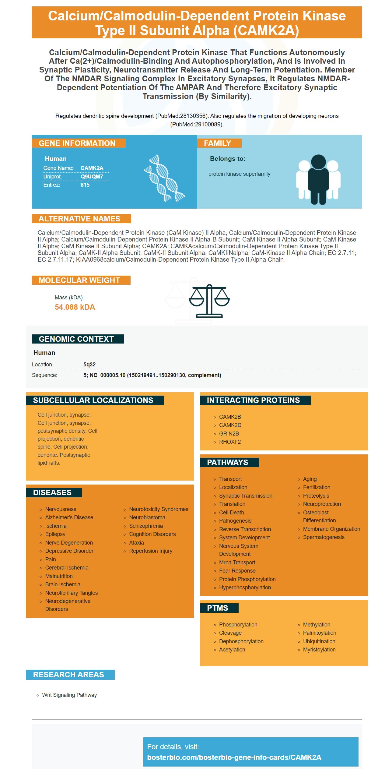
Regulates dendritic spine development (PubMed:28130356). Also regulates the migration of developing neurons (PubMed:29100089).
Human | |
---|---|
Gene Name: | CAMK2A |
Uniprot: | Q9UQM7 |
Entrez: | 815 |
Belongs to: |
---|
protein kinase superfamily |
calcium/calmodulin-dependent protein kinase (CaM kinase) II alpha; calcium/calmodulin-dependent protein kinase II alpha; calcium/calmodulin-dependent protein kinase II alpha-B subunit; CaM kinase II alpha subunit; CaM Kinase II alpha; CaM kinase II subunit alpha; CAMK2A; CAMKAcalcium/calmodulin-dependent protein kinase type II subunit alpha; CaMK-II alpha subunit; CaMK-II subunit alpha; CaMKIINalpha; CaM-kinase II alpha chain; EC 2.7.11; EC 2.7.11.17; KIAA0968calcium/calmodulin-dependent protein kinase type II alpha chain
Mass (kDA):
54.088 kDA
Human | |
---|---|
Location: | 5q32 |
Sequence: | 5; NC_000005.10 (150219491..150290130, complement) |
Cell junction, synapse. Cell junction, synapse, postsynaptic density. Cell projection, dendritic spine. Cell projection, dendrite. Postsynaptic lipid rafts.
Anti-Myeloid-Associated Differentiation (AMD) Marker is an anti-myeloid marker. It is used to determine the expression and localization of a specific protein. Scientists who use this marker have many benefits. Scientists can submit their results for applications, species and other samples. Researchers can earn product credits for their work. The CAMK2A marker is available to scientists from all over the world.
The Anti-Myeloid-Associated Differentiating Marker (A-MADD) is a newly identified protein in Boster Bio. The MYADM sequence is part of the multipass membrane-associated protein family of. The MYADM protein has extracellular domains and is involved in myeloid differentiation. It is a key indicator for diagnosing blood disorders and the progression of lymphoma.
CDDO induces a change in the ratio of transcriptionally active CEBPA to transcriptionally inactive CEBPA which, in turn, causes a change in gene expression. Primary blasts of patients suffering from AML are used to trigger CDDO. The disease is characterized by a lack of differentiation. AML treatment plans may include CDDO therapies.
The results of the study show that the ER control clones differentiate toward neutrophils in the presence of G-CSF+4-hydroxytamoxifen. After four to five days of culture, the C/EBPg ER-expressing Clones were proliferative, but did not differentiate. Results also revealed that C/EBPgER expression doesn't affect the production of B lymphocytes.
Moreover, the A-MYADM antibody inhibits the activity of the SP-A gene, which is a late differentiation-regulator. Both SP-A and anti-MYADM decrease the area under the curve of eosinophils. Additionally, both antibodies reduced the apoptotic phenotypes of eosinophils.
A-MADE2 is a key player in the regulation of transcription factors C/EBPa and G-CSFR. Furthermore, it facilitates differentiation of the initial human AML samples. Both C/EBPa and CEBPG are upregulated in the AML population. The expression of these proteins is highly controlled in the CD150-CMP subset.
When the C/EBPa gene is silenced, the NSC#1 cells that lack the anti-MDA-1 protein cannot replate. In addition, downregulation of C/EBPg reduces the capacity to replate. The cells can then be replated four times. Additionally the CD11b and CD8+ cell distributions are biomarkers that indicate response to anti-MDA-1 treatment. Single-cell RNA sequencing can show the differences between responders or non-responders. In addition, CD45+ cells possess an M2-like transcriptional signature.
The alpha CaMKII promoter normally restricts the expression of transgenes in forebrain areas, however, the original line of EAC3I has higher levels of mRNA in the dorsalstriatum MSNs, possibly due to integration site-dependent effects. However, the expression is weak or absent in the cortex and thalamus. The target protein is expressed in a mosaic-like pattern within the dorsalstriatum. It's also found in 3464% neurons.
The brain expresses four CaMKII isoforms. CaMKIIa, b and CaMKIIg are the most well-known among them, while CaMKIIg is found all over. The expression of CaMKIIa is very high in neurons which account for as much as 11% of protein within some brain regions. CaMKIIb is more prevalent in the cardiovascular system, which includes the heart.
The CAMK2A marker is a neuron-specific serine/threonine kinase which is crucial for synaptic plasticity. The neuronal function is altered in a significant way. could be caused by mutations in this gene. To find out whether this mutation causes greater neuronal activity, researchers conducted an electrophysiological analysis using a multi-electrode array. CAMK2A-mutated h2 embryonic stem cells were more efficient at forming neurons than the corresponding wild-type controls, when contrasted with the controls derived from unrelated embryonic stem cells. They also expressed neuronal markers.
In the past, Chia, Zhong, Niwa and colleagues have identified two siblings suffering from a neurological disease that affects the development of the brain and can trigger frequent seizures. Researchers compared the DNA of the patients to their siblings who were healthy and their parents and then analysed the results. Researchers found that patients had elevated levels of CAMK2A protein, which is a protein vital for memory and learning.
Researchers have discovered that mice homozygous for the CAMK2A gene are viable and show decreased LTP and impaired learning. Heterozygous mice also have significant deficiencies in spatial working memory as well as memories of contextual fear. CAMK2A knockout mice have similar learning issues and impaired LTP. Additionally, mice with neuron-specific mutations in the gene show similar memory and learning impairments.
The CAMK2A gene is extremely preserved from worms to humans. Among humans and worms, the biallelic p.H477Y mutation is pathogenic, and rescue experiments in C. the elegans have suggested that the mutation is the cause of synaptic dysfunction in the worms. These studies also reveal that CAMK2A mutations can be found in both copies the CAMK2A genes.
The CAMK2A gene is mapped to human 5q32 and 18. Justice et al. (2008) discovered that a homologous sequence for CAMK2A was discovered in humans and mice. Human chromosome 18 was later mapped using the CAMK2A gene. The p.H477Y mutation is in the association domain and causes disruption of a histidine residue. In humans, the p.H477Y mutation causes partial loss of function and autosomal recessive inheritance. Heterozygous CAMK2A carriers do not show neuro-developmental symptoms.
To identify synaptic properties that are mature neurons can express CAMK2A. Researchers can determine more mature hpiNs, which have synaptic transmission via NMDAR-mediated synaptic transfer using the CAMK2A marker. These neurons may not be able to differentiate into synapses, but the CAMK2A-progenitor cells continue to mature. They could be contributing to other neurodevelopmental disorders.
PMID: 14722083 by Gaertner T.R., et al. Comparative analyses of the three-dimensional structures and enzymatic properties of alpha, beta, gamma and delta isoforms of Ca2+- calmodulin-dependent protein kinase II.
PMID: 15312654 by Krapivinsky G., et al. SynGAP-MUPP1-CaMKII synaptic complexes regulate p38 MAP kinase activity and NMDA receptor-dependent synaptic AMPA receptor potentiation.
*More publications can be found for each product on its corresponding product page