This website uses cookies to ensure you get the best experience on our website.
- Table of Contents
Facts about Calcium-binding and coiled-coil domain-containing protein 2.
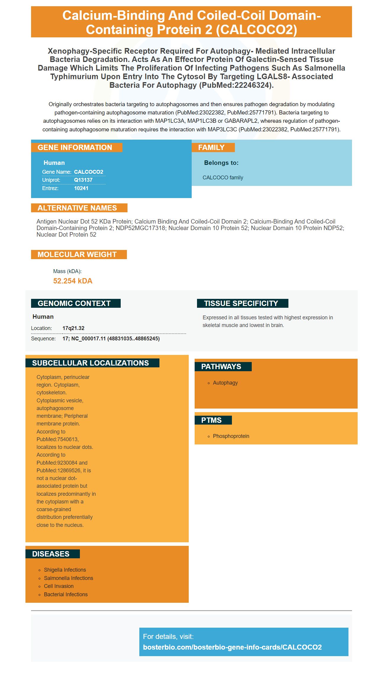
Originally orchestrates bacteria targeting to autophagosomes and then ensures pathogen degradation by modulating pathogen-containing autophagosome maturation (PubMed:23022382, PubMed:25771791). Bacteria targeting to autophagosomes relies on its interaction with MAP1LC3A, MAP1LC3B or GABARAPL2, whereas regulation of pathogen-containing autophagosome maturation requires the interaction with MAP3LC3C (PubMed:23022382, PubMed:25771791).
Human | |
---|---|
Gene Name: | CALCOCO2 |
Uniprot: | Q13137 |
Entrez: | 10241 |
Belongs to: |
---|
CALCOCO family |
Antigen nuclear dot 52 kDa protein; calcium binding and coiled-coil domain 2; calcium-binding and coiled-coil domain-containing protein 2; NDP52MGC17318; Nuclear domain 10 protein 52; Nuclear domain 10 protein NDP52; Nuclear dot protein 52
Mass (kDA):
52.254 kDA
Human | |
---|---|
Location: | 17q21.32 |
Sequence: | 17; NC_000017.11 (48831035..48865245) |
Expressed in all tissues tested with highest expression in skeletal muscle and lowest in brain.
Cytoplasm, perinuclear region. Cytoplasm, cytoskeleton. Cytoplasmic vesicle, autophagosome membrane; Peripheral membrane protein. According to PubMed:7540613, localizes to nuclear dots. According to PubMed:9230084 and PubMed:12869526, it is not a nuclear dot-associated protein but localizes predominantly in the cytoplasm with a coarse-grained distribution preferentially close to the nucleus.
The CALCOCO2 marker is among the most widely-used methods for detecting carbon dioxide in tissues, is one of the most sought-after. It has a low background level and can detect antigens at levels of pg/ml. To learn more about the CALCOCO2 marker and its greatest uses, read on! You can also look up this Boster Bio article
The CALCOCO2 marker is one of the most recent proteins in the human cancer arsenal. Its interactions with IkB kinase and the NFKB pathway could explain the way it can affect human tumors. Additionally, CALCOCO2 is associated with autophagosome assembly and nucleophagy. These interactions may also help researchers understand the evolution of PCa.
CALCOCO2 is a constituent of the nuclear domain 10 (ND10) bodies that are a series of 10 dots per nucleus. They may be connected to the nucleus matrix. Herpes simplex virus-1 infection removes nuclear domain 10 proteins. It is believed that ND10 proteins play a significant role during viral life cycles. It also interacts with GEMIN4, and could be crucial for the formation of ruffles and cytoskeleton organization of actin. The aim of your study will determine the required dilution.
Scientists have outlined the most effective uses of the CALCO2 marker. These researchers include Fostel, Jennifer, and colleagues from the Royal Society of Chemistry: Lord, Phillip, and Hahn, Michael. They also refer to Matondo, Jose M., as well as de la Rosa-Trevin (Jose). Amazon also has Patwardhan, Vikram and Boster Bio, Best Uses for the CALCOCO2 marker.
The pg/ml limit is considered to be the threshold for detection in ELISA methods. Antibodies must be present in the same amount to the antigen that is being identified. Samples with high levels of antigen will be more likely to contain more antibody-antigen complexes and less free antigen. The second step in ELISA analysis is to add the enzyme-conjugated secondary antibody to the sample.
ELISAs are one of the most sensitive immunoassays available. They can be sensitive to as little as 0.1 Fmol, or as sensitive to 10 pg/ml. This depends on the type and interaction between the antigen and the antibody. Indirect detection can increase specificity but it is not always the best results.
ELISA MAX(tm) Standard Sets include all the basic elements required for an ELISA. These kits include a Recombinant protein standard, capture antibody and detection antibody. These kits are a great solution for those with a limited budget. They also come with additional buffers. The ELISA MAX(tm) Standard Sets are perfect for those who have a tight budget. They contain all the components required to conduct a successful ELISA.
The NP ELISA was developed during the SARS epidemic in 2002-2004. Its analytical detection limit for SARS-CoV NP is 50 pg/ml. The diagnostic cutoff was raised to 100 pg/ml , which increases the sensitivity to 95 percent. If it is used in the early stages following the first symptoms of SARS, the NP ELISA is an excellent choice.
ELISA kits are beneficial for research in a variety of fields. For example some researchers employ sandwich ELISA to determine the amount of human TNFa. This technique employs the 2.5X serial dilution (75 pg/ml) of recombinant human thyroid hormone to create an atypical curve. The sandwich ELISA plate is coated with a purified antibody to capture. The samples were then incubated at room temperature for the duration of an overnight.
ELISA kits are indispensable tools for clinical diagnostics and research into human illnesses. The method's ability to detect antigens in the sample will determine its accuracy. True positive results ranged between 10 to 1,377 pg/ml. Low-sensitivity ELISA kits have a lower background. The enzyme present in the sample determines the level of sensitivity of the ELISA kit.
The PIFA assay is a blood test that can be performed for human blood, was developed in the hope of increasing the LOD of an ELISA kit by up to 10 times. The PIFA assay can also be used for research into human cancer. Its slope is slightly higher than that of the PIFA assay. Because of non-specific interactions among proteins in plasma the recovery rate is lower at lower concentrations.
ELISA kits are used to study low-level biomarkers of proteins. It works similarly to real-time PCR but uses o-phenylenediamine dimochloride (oPD) which is a photosensitizer. This method is widely used and is based on the principle that photons amplify the signal.
PMID: 7540613 by Korioth F., et al. Molecular characterization of NDP52, a novel protein of the nuclear domain 10, which is redistributed upon virus infection and interferon treatment.
PMID: 9230084 by Sternsdorf T., et al. Cellular localization, expression, and structure of the nuclear dot protein 52.