This website uses cookies to ensure you get the best experience on our website.
- Table of Contents
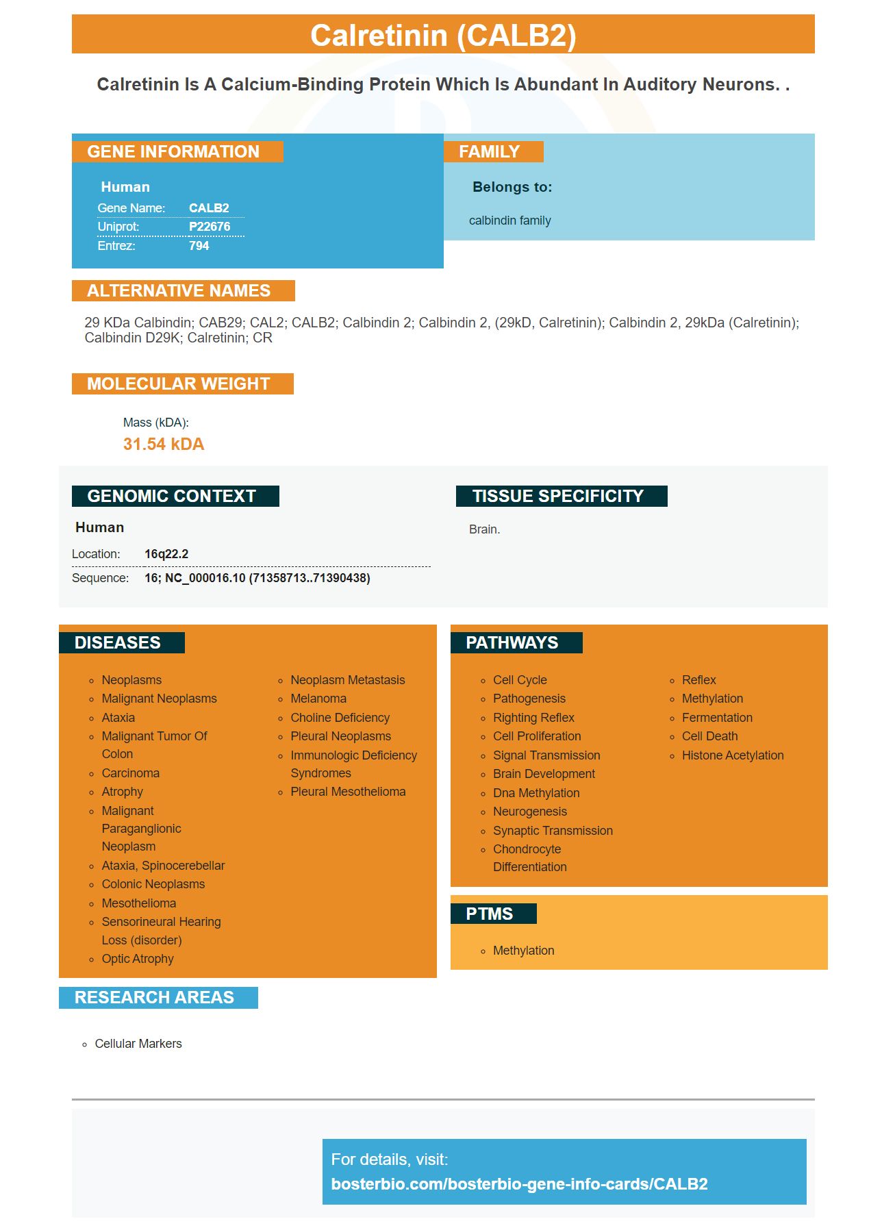


Facts about Calretinin.

| Human | |
|---|---|
| Gene Name: | CALB2 |
| Uniprot: | P22676 |
| Entrez: | 794 |

| Belongs to: |
|---|
| calbindin family |

29 kDa calbindin; CAB29; CAL2; CALB2; calbindin 2; calbindin 2, (29kD, calretinin); calbindin 2, 29kDa (calretinin); calbindin D29K; Calretinin; CR
Mass (kDA):
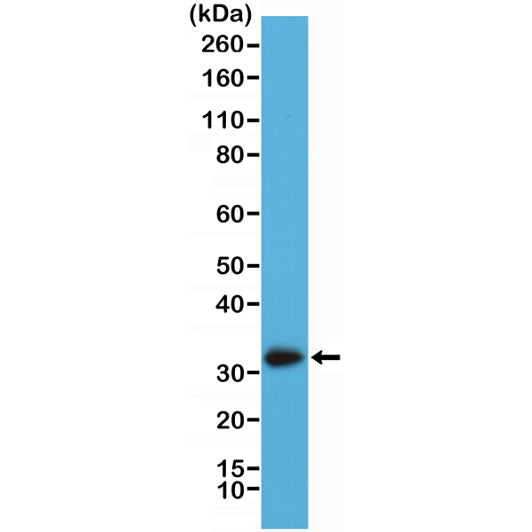
31.54 kDA

| Human | |
|---|---|
| Location: | 16q22.2 |
| Sequence: | 16; NC_000016.10 (71358713..71390438) |
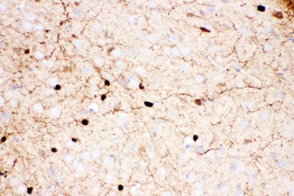
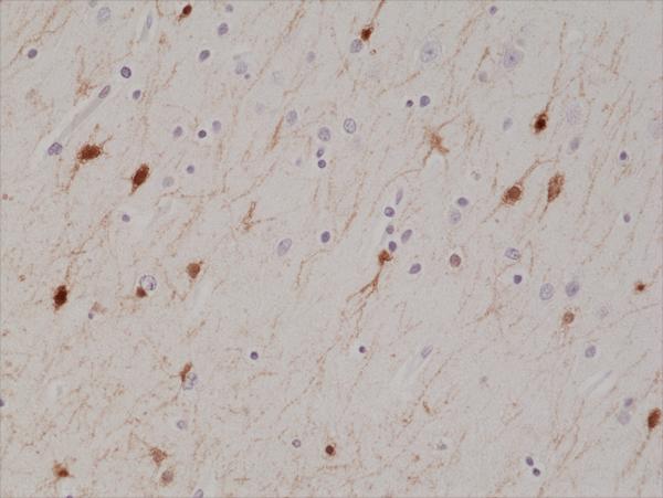
Brain.


PMID: 2618861 by Parmentier M.; The human calbindins: cDNA and gene cloning.
PMID: 2001709 by Parmentier M., et al. Structure of the human brain calcium-binding protein calretinin and its expression in bacteria.