This website uses cookies to ensure you get the best experience on our website.
- Table of Contents
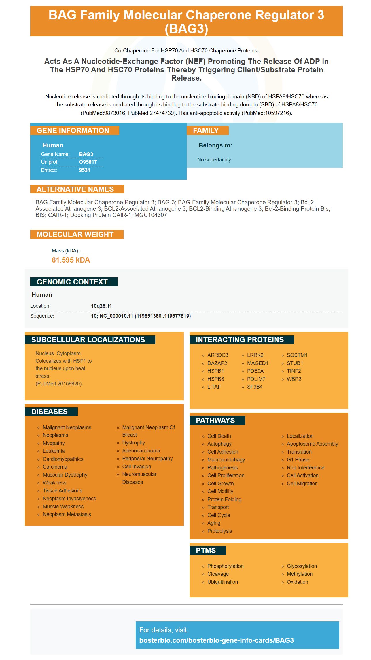
Facts about BAG family molecular chaperone regulator 3.
Co-chaperone for HSP70 and HSC70 chaperone proteins.
Acts as a nucleotide-exchange factor (NEF) promoting the release of ADP in the HSP70 and HSC70 proteins thereby triggering client/substrate protein release.Nucleotide release is mediated through its binding to the nucleotide-binding domain (NBD) of HSPA8/HSC70 where as the substrate release is mediated through its binding to the substrate-binding domain (SBD) of HSPA8/HSC70 (PubMed:9873016, PubMed:27474739). Has anti-apoptotic activity (PubMed:10597216).
Human | |
---|---|
Gene Name: | BAG3 |
Uniprot: | O95817 |
Entrez: | 9531 |
Belongs to: |
---|
No superfamily |
BAG family molecular chaperone regulator 3; BAG-3; BAG-family molecular chaperone regulator-3; Bcl-2-associated athanogene 3; BCL2-associated athanogene 3; BCL2-binding athanogene 3; Bcl-2-binding protein Bis; BIS; CAIR-1; Docking protein CAIR-1; MGC104307
Mass (kDA):
61.595 kDA
Human | |
---|---|
Location: | 10q26.11 |
Sequence: | 10; NC_000010.11 (119651380..119677819) |
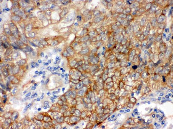
Nucleus. Cytoplasm. Colocalizes with HSF1 to the nucleus upon heat stress (PubMed:26159920).
The BAG3 biomarker has many applications in research. This reagent can be utilized by scientists from all over the world to publish results of species or applications. Scientists may be eligible for product credits. This article will cover the best uses of this reagent. In addition, we'll discuss the mechanisms behind its action. Toll-like receptors (TLRs) are responsible for regulating the function of the BAG3 biomarker.
Ang II significantly increased VMSC proliferative capacity by increasing BAG3 expression. These results suggest that BAG3 is a crucial regulator of Ang II's proliferative activity. Ang II is an inflammatory cytokine with potent potency, that plays a crucial role in atherosclerosis. Toll-like receptor 4 and nuclear factor (NF)-kB pathways are involved in BAG3 upregulation.
The brief exposure to Ang II also accelerated the development of cardiac hypertrophy and damage to the aortic valve. After AngII treatment, blood pressure normalized and the expression of the ICAM1 gene maintained. Cell proliferation and infiltration by CD45+ cells were two of the inflammation markers that were linked to the vascular remodeling induced by AngII. This could suggest that BAG3 could play a role in the prevention and treatment of heart disease.
To test the effects of Ang II on BAG3 expression The mice in the study group were treated with angII. In the presence of Ang II, HTR-8/SVneo cells produced Ang 1-7. The Ang II+2W group produced Ang 1-7. Incubations with no Ang II also produced Ang 1-7. These results indicate that Ang II induces VSMC proliferation in a dose-dependent manner.
The measurements of blood pressure were also taken longitudinally in mice in the Ang II-treated group. The AngII group had significant increases in the total heart/tibial length and left ventricular/body weight at the time of baseline. These changes in phenotypes maintained over time, which suggests that BAG3 plays a significant role in the prevention and treatment of cardiovascular disease. The study also employed littermates of the same age as controls.
The AT1R knockdown caused by Ang II slowed tumor growth and invasion in HepG2 cells. Furthermore, mice treated with Ang II had increased BAG3 expression, which indicates that these two drugs reduce the growth of tumors and their invasion in a biphasic fashion. These results don't support the existence of a synergistic link between Ang II and AT1R. Inhibiting the effects of AT1R on HCC cells is a possibility.
The AT2R knockdown that Ang II induces inhibited the glucose transporter 2 and insulin. Additionally, it decreased the apoptosis caused by cAMP. However, AT2R knockdown also suppressed RAS genes and induced angiotensin-(1-7)-independent G1-phase cell cycle arrest. It also decreased ERK and pAkt expression.
Ang II treatment caused an increase in VMSC proliferation. This was related to an increase in MMP2 (and MMP9) expression, which are genes that control VSMC proliferation and migration. The invasive behavior of AngII stimulated VSMCs was linked to an increase in MMP expression. The effect of SFN on HVSMC proliferation and migration was reversed when CHD1L was silenced.
We aren't sure of the mechanism by which the Ang II-induced VSMC growth. Although Ang II is a key vasoactive peptide, it plays important roles in pathogenesis of cardiovascular diseases. Ang II induces plaque vulnerability by causing rapid growth and movement of VSMCs. In Ang II, cAMP/PKA is stimulated. signaling pathway, ERK1/2, and the NADPH oxidase subunit. Furthermore, it induces the proliferation of HVSMCs.
The activation of STAT3 leads to translocation into the nucleus. Ang II treatment increased the amount of STAT3 in VSMCs. GSTpi blocks Ang II-induced STAT3 nuclear translocation. Ang II treatment enhanced VMSC proliferative capacity. By blocking Ang II-induced translocation, GSTpi inhibits VMSC migration. Transwell assays were employed to further investigate the effect of GSTpi inhibition on VSMCs.
Ang II treatment increased VMSC growth and migration in vitro. This was also evident after GSTpi treatment. The treatment slowed atherosclerosis. The Ang II drug stimulates the phosphorylation of STAT3 at tyrosine 705 and stimulates STAT3 transfer to the nucleus. STAT3 plays a key role in regulating the proliferation and migration of VSMCs.
CHD1L is a negative regulator of VMSC proliferative. CHD1L knockdown also restored the effects of AngII in VMSC expansion and migration. CHD1L inhibition prevented AngII-induced VMSC expansion in VSMCs. The study also revealed that FOXO3a knockdown reversed the effects of CHD1L deficiency on AngII-induced VSMCs.
VSMCs were transfected using plasmids, and then cultured in 6-well plates for 36 h and then incubated in NG for 24 h. The treatment with Ang II boosted S-glutathionylation of STAT3 in VSMCs, regardless of whether GSTpi is expressed in excess. It is noteworthy to note that GSTpi is not affected by the effects of Ang II on STAT3 in VSMCs.
CCK-8 assay that analyzes the cellular conversion of colorimetric reagents, was used to determine whether Ang II increased VMSC proliferation. In this study, Ang II treatment was discovered to increase VMSC proliferative capacity, whereas SFN had no effect. The increase in cellular viability was associated with reduction in the amount of SFN. This study should not be taken to prove that Ang II is effective in treating VMSC.
Steven Boster has created the Boster Bio: Best Uses for the BAG3 Marker. He is known as "he who turns science into a toilet." He began his career in 1993 by creating a variety of products for IHC. Then, he expanded his business to become one of China's biggest catalog antibody companies. PicoKine(tm) was also developed by his company. Additionally, he has incorporated trade secrets that were proprietary to him into this platform to provide high-sensitivity ELISA kits.
Through the TLR4/NFK p65 signaling pathway, Ang II regulates BAG3 transcription. In fact, the BAG3 signaling pathway is critical for the survival of many cancer cells, which includes tumors and normal tissues. The Ang II signaling pathway stimulates VMSCs autophagy, by altering BAG3 transcription. Furthermore, this receptor triggers the autophagy response in cancer cells, and blocks tumor growth.
The BAG3 antibody was examined against negative and positive samples from human cancer tissues. Its ability to bind to antigens has been demonstrated in sporadic forms of Dilated Cardiomyopathy. BAG3 expression is high in certain cells, and is associated with resistance to mechanical stress. BAG3 expression was linked to fibrosis in the model of unilateral obstruction of the ureteral tract in rats. This was in correlation with an increased synthesis of extracellular matrix protein.
BAG3 overexpression in the human brain hinders autophagy by promoting autophagy-inducing protein which includes the LC3II. The levels of other cellular proteins such as GAPDH were not affected by BAG3's overexpression. BAG3 also blocks the activity of the protein-coding enzyme Beclin-1. This suggests that BAG3 overexpression could lead to an autophagy pathway that is dysfunctional.
BAG3 is a great marker for research into biomarkers. It is used in various applications, including immunohistochemistry and cell biology. It is compatible with both primary and secondary antibodies. The best use of the BAG3 Marker is for immunohistochemistry. Additionally, the Boster Bio: Best Uses for the BAG3 Marker
BAG3 is involved in many physiological processes, as well as the immune system. Its antigen binding activity is enhanced in the presence of BAG3 in different tissues, including in cancer cells. This marker is compatible with all types of cells including cancer cells and immune diseases. You can purchase the antibodies used in this biomarker from various online sellers. Be sure that the anti-BAG3 antibody is tested in both mouse and human cells before you purchase it.
Ang II is a major factor in the proliferative responses of VSMCs. It regulates BAG3 expression by upregulating other factors involved in cell proliferation. Boster Bio sells this antibody. Ab63957, the primary antibody, detects BAG3 expression within the HK2 cell line. Secondary antibodies can be used to observe the protein bands.
PMID: 9873016 by Takayama S., et al. An evolutionarily conserved family of Hsp70/Hsc70 molecular chaperone regulators.
PMID: 10597216 by Lee J.H., et al. Bis, a Bcl-2-binding protein that synergizes with Bcl-2 in preventing cell death.