This website uses cookies to ensure you get the best experience on our website.
- Table of Contents
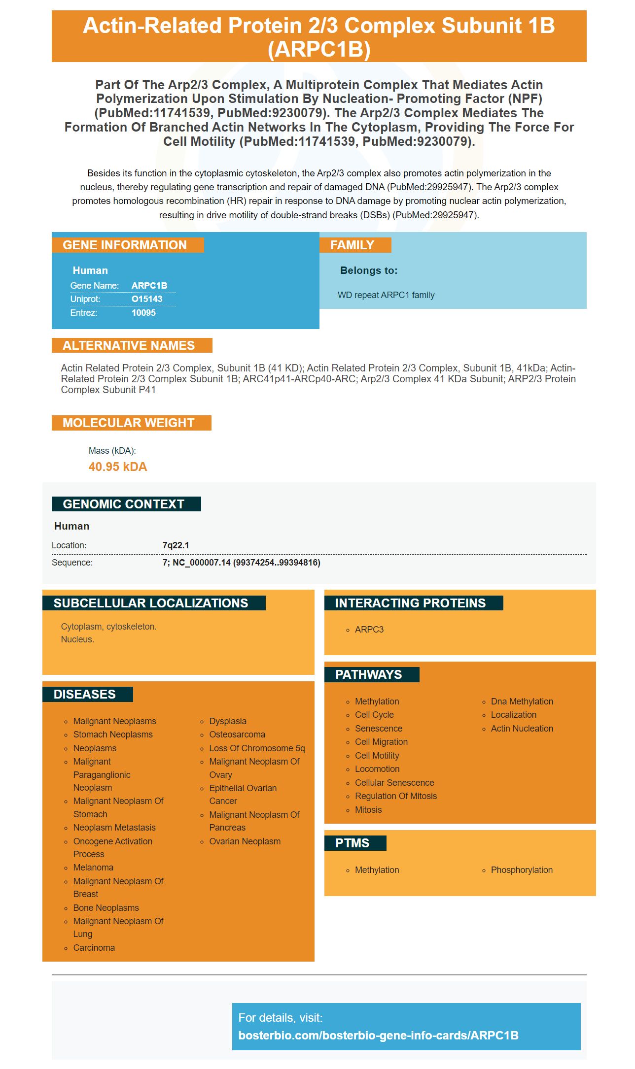
Facts about Actin-related protein 2/3 complex subunit 1B.
Besides its function in the cytoplasmic cytoskeleton, the Arp2/3 complex also promotes actin polymerization in the nucleus, thereby regulating gene transcription and repair of damaged DNA (PubMed:29925947). The Arp2/3 complex promotes homologous recombination (HR) repair in response to DNA damage by promoting nuclear actin polymerization, resulting in drive motility of double-strand breaks (DSBs) (PubMed:29925947).
Human | |
---|---|
Gene Name: | ARPC1B |
Uniprot: | O15143 |
Entrez: | 10095 |
Belongs to: |
---|
WD repeat ARPC1 family |
actin related protein 2/3 complex, subunit 1B (41 kD); actin related protein 2/3 complex, subunit 1B, 41kDa; actin-related protein 2/3 complex subunit 1B; ARC41p41-ARCp40-ARC; Arp2/3 complex 41 kDa subunit; ARP2/3 protein complex subunit p41
Mass (kDA):
40.95 kDA
Human | |
---|---|
Location: | 7q22.1 |
Sequence: | 7; NC_000007.14 (99374254..99394816) |
Cytoplasm, cytoskeleton. Nucleus.
Primary antibodies for ARPC1B are required to identify proteins and detect activity in biological samples. These antibodies are produced by Boster Bio and are well-validated on Western Blotting, Immunohistochemistry, and ELISA. Researchers have used Boster antibodies for decades. They are highly specific. Boster antibodies have been used in many experiments and are highly cited.
A high-affinity Western Blot detection method is one of the best ways for positive results. These methods are similar to those used for other immunoassay technologies, including ELISA. Both rely upon specific antibodies that bind a target antibody and a detection device that measures the amount antigen bound to the membrane. High-affinity antibody coated is an important ingredient in maximizing the assay's sensitiveness. Boster Bio has mastered this method and is trusted by scientists worldwide.
The primary and secondary antibodies should recognize the same antibody to maximize the effectiveness of western blot antigen detection. There are a variety of methods for detecting antibodies on a western blot, and each one has its own advantages. Monoclonal antibodies will only recognize one antigenic epitope. This will increase the specificity while decreasing background. A polyclonal antigen will recognize many antigenic epitopes. This is recommended for more complicated studies.
An ELISA allows you to compare the relative levels and give you a yes/no result. ELISA data is typically graphed using fluorescence vs..concentration. The resulting graph will look like a sigmoidal curve. Boster Bio Picokine ELISA can detect levels down to 1 ng.
The target protein will be detected when the primary antibodies is used. The secondary antibody binds with the target protein and is then incubated using enzyme-conjugated antibodies. This process may require several exposures depending upon the concentration of the primary antibodies. This procedure can take anywhere up to six hours. Depending on how high the target protein is, the test might take longer than one to complete.
The ECL-chemiluminescent detection method uses antibodies conjugated HP and a substrate. The substrate is bound to the enzyme, which then releases excited intermediates that emit strong blue light at 450nm. The enzyme-substrate reacts only to emit light, so the signal output stops once the substrate is gone.
A 50 mM Tris solution (pH 7.4) was used to prepare an adult testis lysate. This solution contained 1% NP-40 as well as protease/phosphatase inhibitor cocktails. The membranes were exposed with an Xray film. It should contain the protein side. The exposure time should be approximately one minute. The light emission peak should occur between five and thirty minutes after the substrate has been reacted with antibody. After the reaction, the nitrocellulose membrane could be transferred to a plate with 96 wells.
All horseradish conjugates can be used with the Clarity Max Substrates. The Clarity Max substrates can be used for all types western blots. They are compatible to most horseradish peroxidase conjugates. They are ideal for all types of western blots. The Clarity Max is compatible with all types horseradish peroxidase conjugates.
Depending upon your application, you can choose from a variety Enhanced Chemical Luminescent Substrates. The Pierce ECL Substrat is recommended for western analysis with high levels of protein. For IHC-P experiments, the HRP-conjugated Super Vision Assay Kit SV0002-1 is used. These reagents have been designed to increase the signal intensity and sensitivity.
Boster Biologicals' ARPC1B chromogenic detector kit is capable of detecting a wide range of proteins and other biomarkers. These kits can be used for immunohistochemical analysis and chromogenic staining. Boster's ARPC1B chromogenic detection system uses NBT nitro-blue tetrazolium and BCIP 5-brotho-4-chloro-3'-indolyphosphate. Picogram-level sensitivity is achieved by the chromogenic detection system.
A new method for western blotting increases the efficiency of proteins. Colloidal gold membrane staining is much more sensitive than standard membrane staining. The resulting bands will appear red against a pink background. They will only be visible for proteins with more than 2 ng per band. A phosphate-buffered-saline (PBS),-treated membrane could produce a stronger signal. Before you start the membrane staining process, make sure to prepare a solution of ferrous sulfurate.
Prepare the transfer buffer. It contains 25 mM Trisbase and 190mM glycine. 20% methanol is added. 4% paraben is added to the solution. The membrane should be left to incubate at 37°C for between 15 and 30 minutes depending on how much protein is present. To remove any antibodies, wash the membrane three more times with TBS Wash Buffer. Next, mix the target protease sample with a solution 4X Dual-Color Protein Loading Bulffer. Mix the BCIP/NBT sample with the BCIP/NBT substrate solution at a ratio of 3:1.
Picokine (tm) increases the sensitivity for ELISA assays up to the picogram-level. Picokine (tm) ELISA Kits have been validated in numerous samples. The validation process and images are available upon request. Supervision, which is a polymer-based secondary antigen that can be used to save time in IHC staining, is available. It is based off of insights into immunogen design. This technique can be improved by Sanbio's comprehensive technical support program.
The nitrocellulose membrane used in the procedure has a high affinity for proteins and is compatible with many detection methods. The nitrocellulose membranes can immobilize proteins, glycoproteins, nucleic acids, and nucleic acids of various molecular weights. The nitrocellulose membranes allow for the detection of both proteins and nucleic substances with one method.
PMID: 9230079 by Welch M.D., et al. The human Arp2/3 complex is composed of evolutionarily conserved subunits and is localized to cellular regions of dynamic actin filament assembly.
PMID: 11741539 by Gournier H., et al. Reconstitution of human Arp2/3 complex reveals critical roles of individual subunits in complex structure and activity.