This website uses cookies to ensure you get the best experience on our website.
- Table of Contents
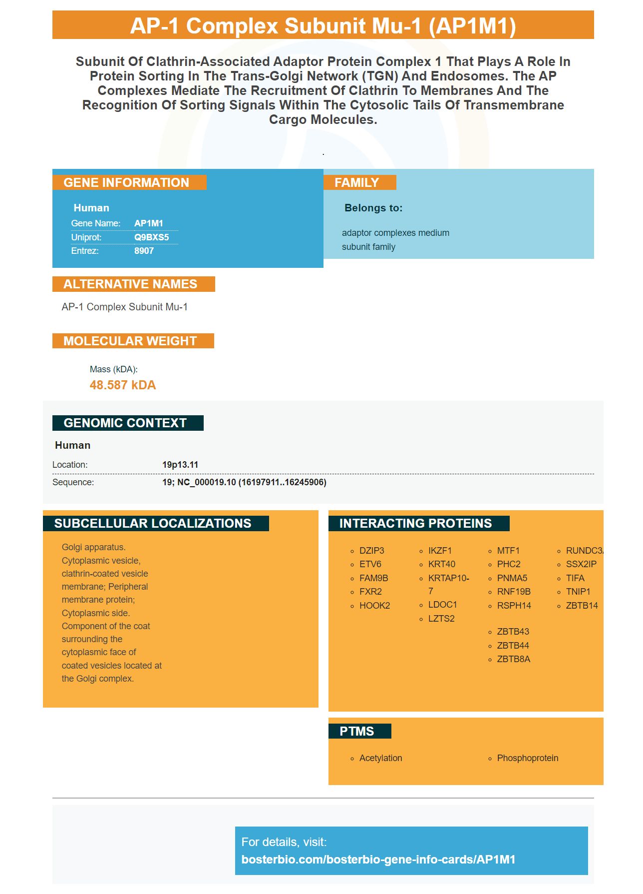
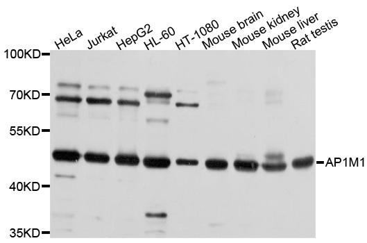
Facts about AP-1 complex subunit mu-1.
.
Human | |
---|---|
Gene Name: | AP1M1 |
Uniprot: | Q9BXS5 |
Entrez: | 8907 |
Belongs to: |
---|
adaptor complexes medium subunit family |
AP-1 complex subunit mu-1
Mass (kDA):
48.587 kDA
Human | |
---|---|
Location: | 19p13.11 |
Sequence: | 19; NC_000019.10 (16197911..16245906) |
Golgi apparatus. Cytoplasmic vesicle, clathrin-coated vesicle membrane; Peripheral membrane protein; Cytoplasmic side. Component of the coat surrounding the cytoplasmic face of coated vesicles located at the Golgi complex.
You might be wondering: What is the AP1M1 gene and how does it work? You'll find out more about the AP1M1 marker and its clinical applications in this article. You can also learn more information about the AP1M1 combination with other markers of tumor stem cells. But before we get into that, let's quickly recap some basics.
The AP1M1 marker is a candidate for protein sorting within the trans-Golgi system. This bipartite protein is part of the AP complexes family and plays a role in the sorting of proteins at the transmembrane space. AP complexes mediate clathrin recruitment to membranes. They also recognize sorting signals found in the cytosolic trail of transmembrane freight molecules.
The AP1M1 marker can be used in a variety of ways in plant research. In plants, AP1M1 is involved in vesicular transport, which involves moving protein from one organelle to another. AP1M1, which is required for early senescence, is involved in vesicular delivery in rice.
The expression of other RTKs requires the AP1M1 gene protein. HCC cells express ErbB4 as well as MET, which have been shown to be induced in AP-1 cells. Although AP-1 was not directly quantified by histological studies it is believed that it supports the expression other RTKs such EGFR. It is known that the AP-1 complex supports the expression of other RTKs, such as EGFR and HER2.
Higher eukaryotes have m1A which is fatal to embryos. In plants, AP1M2 is an intermediate-sized subunit of the AP-1 complex. Higher eukaryotes also have similar levels of expression for m1A. Furthermore, a single mutant of AP1M2 was found to grow poorly despite the presence of an external epitope. These results suggest that the function of m1A in Arabidopsis can be confirmed.
The AP1M1 gene marker is useful in genetic studies of plant cells. The AP1M2 marker is useful for genetic studies of plant cells. A double mutant of ap1m1 is almost deadly. Despite its high functionality and usefulness, the AP1M1 mark is not widely used by humans.
A subset if paragangliomas has been found to express immunohistochemically tumor stem cell markers. These markers could serve as therapeutic targets in cancer cells or predictive indicators of malignancy. Although the exact mechanisms behind their expression are not known, they seem to correlate with tumor size or genetic background. Below are some examples of markers which may be co-expressed and other tumor stem cell markers.
TMA series TMA2 and TMA28 have been found to co-express two markers, SOX2 as well as LIN28. The co-expression of SOX2 (and LIN28) is consistent with their shared regulatory function and is in line with the known functions these two proteins. SOX2 acts directly as a binding partner to LIN28A, regulating ESC activity. SOX2 and LIN28A are also co-expressed in PCC/PGL samples.
DCLK1 and NESTIN also exist in tumor stem cell, but at lower levels compared to unsorted iCCAs. Seven candidate markers for tumor stem cells were identified using a tissue microarray. DCLK1 was highly expressed in pCCACD90+ cells. NESTIN, EpCAM, and NESTIN were not significantly different among sorted HT29 cell lines.
Intact tissue gene expression is determined based primarily on the number of cells in the area and their anatomical distribution. Different cell types produce different signal-to noise ratios. Schwann cells, for example, are rare tumor stem cell markers and are co-expressed with a large number of genes. These genes are associated with cancer stem cells and are useful in tumor diagnosis. This news is very exciting for cancer patients, and could be a positive step forward in the field.
KIFC1 is another potential marker for CSC. It has been shown in HCC to induce epithelial-mesenchymal transformation and subsequently to promote metastasis. Westphalen and his colleagues also found that DCLK1 is a hallmark in CSCs in colon carcinoma. This study revealed a novel link between DCLK1 tumor cells and CSCs in perihilar CCA.
The results suggest that cancer stem cells may play a role when it comes to drug resistance. The study showed that LUAD patients with TCGA tumour tissues had higher mRNA expression based tumor stemness than the control group. The mRNAsi index was significantly associated with clinical features. The blue module was deemed the most pertinent, while the yellow module contained 50 important genes. These genes are enriched for cell cycle, chromosome segmentation, and chromosome area. Six hub genes have been identified using the protein–protein interaction network and Molecular Complex Detection.
Modules A-F genes were found to be enriched in regions distal to epithelial niches and in the mesenchyme. This suggests that these genes are correlated in their cellular abundance across the different regions. The dissection would also show a greater number of epithelial cells.
A methylation-induced cell response regulates gene expression without altering DNA sequences. It alters DNA stability, conformation, and interactions between DNA, proteins. The process is mediated by DNA methyltransferases, which add methyl groups to DNA at the 5' carbon position of the cytosine ring. Currently, AP1M1 expression in tumors with GBM or LGG is elevated.
Expression of AP1M2 is correlated with immune invasion in fourteen tumor types. It also correlates to 16 cancers CD4+ T cell infiltration. Moreover, it correlates with the presence of DC, macrophages, and neutrophils. These findings indicate that AP1M2 is a major factor in the development BRCA. It could play an important role in early diagnosis and treatment of the disease.
AP-1 is a tumor suppressor that is involved in the maintenance of cell proliferation in various types of cancer. For membrane recruitment, AP-1 is required along with a subset accessory proteins. These accessory proteins should be further studied in human tumors. AP-1GGA2 might block multiple cancer-related signaling pathways. However its target is still unknown.
The AP1M1M1M1 gene complex forms an integral part in the AP-1 proteins complex. It is an essential component in the clathrin adsorbor AP-1. It regulates the formation of clathrin-coated pits, targets Rab5 isoforms, and induces membrane scission. It is also required for early endosome formation.
The new technique of proteomics-based proteomics has many clinical applications, including the identification and validation of therapeutic targets. MS technology is used to extract proteins from biological samples. Peptides are then digested and separated by liquidchromatography with MS analysis. A typical mass spectrometry-based proteomics test will yield a protein library that contains approximately 2,000 proteins and/or peptides.
PMID: 18073204 by Wonderlich E.R., et al. The tyrosine binding pocket in the adaptor protein 1 (AP-1) mu1 subunit is necessary for Nef to recruit AP-1 to the major histocompatibility complex class I cytoplasmic tail.