This website uses cookies to ensure you get the best experience on our website.
- Table of Contents
Western blotting (also called Protein Immunoblotting) is an analytical technique used to detect specific proteins in the given sample. It uses SDS-polyacrylamide gel electrophoresis (SDS-PAGE) to separate various proteins contained in the sample. The separated proteins are then transferred or blotted onto a matrix, where they are stained with antibodies specific to the target protein. Expression details of the target proteins in the given cells or tissue homogenate can then be obtained through analyzing the location and intensity of the specific reaction. Western blotting analysis can detect target protein as low as 1 ng due to high resolution of the gel electrophoresis and strong specificity and high sensitivity of the immunoassay. This method is used in the fields of molecular biology, biochemistry, immunogenetics and other molecular biology disciplines for various experiments.
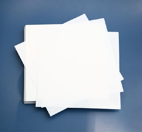
Filter paper (or blotting paper) is important to ensure quick and efficient transfer of molecules from the electrophoresis gel to the matrix membrane. It is used for transfer sandwiches and cassettes during Western blotting and assists with transferring proteins unto polyvinylidene fluoride (PVDF), nitrocellulose, and other types of membranes. Western blot filter paper is a semi-permeable paper barrier used to separate fine solid particles from liquids or gases. It can be purchased as pre-cut cotton fiber sheets for wet, semi-dry, passive, or electrophoretic transfer of proteins. The thickness of the paper determines the flow rate of the protein or nucleic acid.
During the process of western blotting, Mahmood et al., 2012 described the steps of electrotransfer as follows:
*Ensure there are no air bubbles between the gel and membrane and squeeze out extra liquid.
*The running time should be proportional to the thickness of the gel, so this may be reduced to 45 minutes for 0.75 mm gels.
It is possible to reuse the filter papers after initial use, as long as they have been dried at the cleaning area. However, they do not last forever and remembering to switch them out is important for your experiments. A common source of transfer issues occur due to the tightness of the sandwich. This can be reduced by replacing your sponge and filter papers regularly. In particular with the sponges, using extra sheets of filter paper to compensate for the sponges that are worn out may lead to inconsistencies with the transfers. This can be detected due to decreased signals and the unfortunate disappearance of the proteins.

Boster Bio specializes in manufacturing primary antibodies for WB, IHC, and ICC/IF as well as producing high quality ELISA and reagent kits. Our lab runs hundreds of WB every week for our internal production quality control, and we use the reagents we manufacture ourselves for our ELISA kits and IHC- and WB-validated antibodies, so we ensure all our extraction kits, buffers, controls, solutions, etc. will help you obtain clean and specific results for accurate localization and identification of antigens.
We provide western blotting filter paper that is 0.158 mm thick with 2 sizes: 12.5 cm x 12.5 cm (Catalog # AR0172) and 9 cm x 7.5 cm (Catalog # AR0173). As described, Boster’s filter paper is pre-cut cotton fiber that works with wet, semi-dry, passive, or electrophoretic transfer of proteins from SDS-PAGE gel to PVDF, nitrocellulose, and other membranes. Our western blot filter papers are manufactured with ultrapure water and contain no additives that can interfere with any application. These filter papers are smooth and suitable for use with alcohol and other organic solvents involved in protein transfer and nucleic acid blotting. They also allow for a uniform flow of buffer through the gel to the transfer membrane in a blotting sandwich. The filter papers are pre-cut in several sizes for use with most mini gel sizes, tank transfer cassettes, and semi-dry blotters, creating ease for use. They have also been tested and recommended for use with various protein methods, including tank and semi-dry transfer.
There are five main steps involved in the western blot workflow, described as follows:
Unlock the true potential of your research and obtain invaluable insights from your samples with our exceptional service. Contact us today to discuss your project requirements and experience the transformative capabilities of our professional Western blotting service.