This website uses cookies to ensure you get the best experience on our website.
- Table of Contents
Western blotting (also called Protein Immunoblotting) is an analytical technique used to detect specific proteins in the given sample. It uses SDS-polyacrylamide gel electrophoresis (SDS-PAGE) to separate various proteins contained in the sample. The separated proteins are then transferred or blotted onto a matrix, where they are stained with antibodies specific to the target protein. Expression details of the target proteins in the given cells or tissue homogenate can then be obtained through analyzing the location and intensity of the specific reaction. Western blotting analysis can detect target protein as low as 1 ng due to high resolution of the gel electrophoresis and strong specificity and high sensitivity of the immunoassay. This method is used in the fields of molecular biology, biochemistry, immunogenetics and other molecular biology disciplines for various experiments.
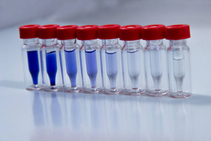
Enhanced Chemiluminescence (ECL) Western Blot Substrate is a very sensitive, non-radioactive, enhanced luminol-based chemiluminescent substrate that allows for easy detection of horseradish peroxidase (HRP) on immunoblots. HRP is a common molecule conjugated to antibodies. ECL Western Blot Substrate has the capability of offering an optimal signal to visualize proteins marked by HRP, with a clear background for easier interpretation. Studies have indicated these are some of the important factors needed to maximize your western blotting results. The luminescence is produced due to the HRP-mediated reaction with the substrate and is indicative of the amount of HRP-labeled antibodies. Furthermore, a higher amount of HRP-labelled antibodies produces more visible chemiluminescence, which also allows for the measurement of the target protein indirectly. This chemiluminescence can be visualized using charge-coupled device (CCD) image systems or x-ray film. It can be used in combination with nitrocellulose or polyvinylidene fluoride (PVDF) membrane, as well as multiple different blocking buffers. Beyond western blots, ECL Western Blot Substrate can also be used for immunoblots, dot blots, and any type of blotting application that uses HRP-conjugates.
Colorimetric detection is an alternative to chemiluminescence detection, from triggering a reaction between the enzyme conjugated to the second antibody and the substrate to produce a colored precipitate. The pros and cons are described in the following table:
| Detection type | Advantages | Disadvantages | Appropriate setting for use |
|---|---|---|---|
| Colorimetric | Fast, cheap, and no special equipment required | Not very sensitive and lots of protein required | A quick and simple test of presence or absence of a protein |
| Chemiluminescence | High sensitivity and best for low expressed proteins | Requires specialist equipment and multiplexing is difficult | Determining quantity of a small amount of protein |
The following is a simplified overview of the assay protocol:
Note: The membrane must be thoroughly washed after incubation with the HRP conjugate.
Note: For CCD detection, put the blot membrane in the CCD and detect the chemiluminescence image according to the manufacturers’ instructions.
Boster Bio specializes in manufacturing primary antibodies for WB, IHC, and ICC/IF as well as producing high quality ELISA and reagent kits. Our lab runs hundreds of WB every week for our internal production quality control, and we use the reagents and kits we manufacture ourselves for our ELISA kits and IHC- and WB-validated antibodies, so we ensure all our extraction kits, buffers, controls, solutions, etc. will help you obtain clean and specific results for accurate localization and identification of antigens.
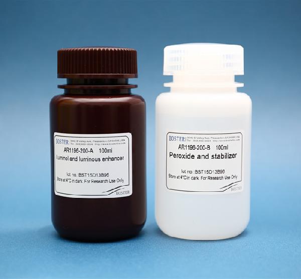
We provide ECL Plus Western Blotting Substrate (Catalog # AR1196-200), which is an ultra-sensitive luminol-based chemiluminescent substrate appropriate for the detection of HRP at high sensitivity levels. Boster’s ECL Plus Western Blotting Substrate has a sensitivity level of low picogram to mid-femtogram As described earlier, our substrate is suitable for any blotting application that uses HRP-conjugates. It can be used with many different blocking buffers and on nitrocellulose or PVDF membranes with low background produced. The product is shipped as Reagent A (luminol and luminous enhancer) and Reagent B (Peroxide stabilizer), which are ready-to-use 1x solutions.
The additional materials required for your experiment include the following:
Make the most of our exceptional service to expedite your research and uncover valuable insights from your samples. Contact us today to discuss your project requirements and witness the transformative results of our industry leading Western blotting service.