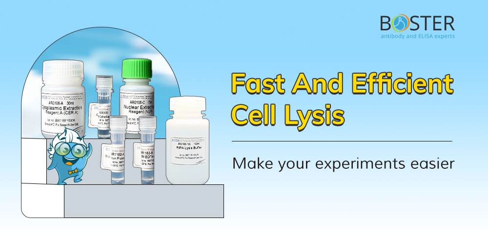
Cell Lysis: A Simple Guide

1. Definition

Cell lysis refers to the process of breaking down the cell membrane through physical, chemical, or enzymatic methods, allowing the release of intracellular components such as proteins, nucleic acids, and small molecules. Cell lysis is crucial in molecular biology and biochemistry, especially when extracting and analyzing intracellular target molecules.

2. Cell lysis method

  • Mechanical/Physical Methods:
  • Ultrasonication: Uses high-frequency sound waves to generate powerful shear forces through cavitation, breaking the cell membrane.
  • Bead Beating: Cell suspensions are mixed with tiny grinding beads and agitated at high speed, causing cells to break from collisions.
  • High-Pressure Homogenization: Suspensions are forced through a high-pressure needle valve, then impacted on a collision ring and released into a low-pressure environment.
  • Cell Physical Methods:
  • Osmotic Shock: Cells are placed in a hypertonic environment and then quickly moved to a hypotonic environment, causing them to swell and burst.
  • Freeze-Thaw Cycles: Cells are repeatedly frozen and thawed, utilizing the mechanical disruption from ice crystal formation and dissolution to break the membrane.
  • Chemical Methods:
  • Detergent Lysis: Lipophilic or anionic detergents dissolve the lipid bilayer of the cell membrane while preserving the integrity of intracellular structures.
    • Enzymatic Methods:
    1. Lysozyme Treatment: Used to lyse bacterial cell walls, often combined with other methods
    2. Proteases and Nucleases: Applied after lysis to selectively degrade or remove specific cellular components such as proteins or nucleic acids.
    3. During cell lysis, controlling temperature, timing, and using enzyme inhibitors is crucial to prevent protein degradation and functional loss. The choice of lysis method may affect the conformation or function of target proteins, so the method must be selected based on experimental needs.

3. Chemical Lysis Buffers Components

  • Detergents: Break down cell membranes; different detergents are suited for extracting different protein types
  • Buffers: Provide a stable pH environment to maintain protein structure and function.
  • Salts: Maintain physiological ionic strength, stabilize protein structure, and prevent non-specific protein binding.
  • Chelating Agents: Bind metal ions, inhibiting metal-dependent proteases and protecting proteins from degradation.
  • Protease Inhibitors: Prevent proteins from being degraded by proteases during lysis.
  • Phosphatase Inhibitors: Protect the phosphorylation state of proteins, preventing the removal of phosphate groups.

    Taking the commonly used RIPA lysis buffer as an example, below is its formulation:
        50mM Tris•HCl pH 7.6
        150mM NaCl
      1% NP-40
      0.5% sodium deoxycholate
      0.1% SDS
    Different lysis buffer components work together to effectively lyse cells, extract target proteins, and stabilize these molecules. Choosing the appropriate lysis buffer components and combinations for different cell types can enhance protein extraction efficiency while preserving the natural structure and function of proteins.

4. How to choose different types of lysis buffer

To extract proteins from different cellular locations, different types of cell lysis buffers are required due to the distinct structures and compositions of various cellular regions. Different lysis buffer formulations can selectively lyse specific cellular structures, thereby releasing proteins from the target locations.

For example, the Enhanced RIPA Lysis Buffers have advantage ranges in the following areas:

  • Membrane Protein Extraction: Enhanced lysis buffers provide stronger lysis capability when studying membrane or other insoluble proteins.
  • Complex Samples with Multiple Proteins: For challenging tissue samples or samples rich in organelles, enhanced lysis buffers can thoroughly lyse organelle membranes, releasing all components for downstream analysis.
  • Experiments Requiring Efficient Lysis: Enhanced lysis buffers are the preferred choice when a complete lysis of the sample and extraction of all protein types is needed. Enhanced RIPA, for example, is ideal for extracting hydrophobic membrane proteins or other difficult-to-solubilize proteins due to its effective membrane-disrupting components.
Here are the commonly used types of lysis buffers for extracting proteins from different cellular locations.

Location of ProteinLysis Buffer Recommended
Whole CellNP40、RIPA
CytoplasmicCytoplasmic and Nuclear Protein Extraction Kit
Membrane boundNP40/RIPA
NuclearCytoplasmic and Nuclear Protein Extraction Kit
MitochondriaRIPA
Golgi ApparatusEnhanced RIPA


5. Procedure for Lysis of Monolayer-cultured Adherent Mammalian Cells

(use RIPA as an example)

Note: Pre-chill an appropriate volume of RIPA Lysis Buffer at 4°C. If desired, add protease inhibitor and phosphatase inhibitor to the lysis buffer immediately before use.

  • Harvest

    1. In a microcentrifuge tube, resuspend 5×10⁶ cells in the growth media by scraping the cells off the surface of the plate with a cell scraper. Centrifuge harvested cell suspension at 600xg for 5min, then carefully remove and discard the supernatant.

  • Wash

    2. Resuspend the cells in chilled PBS. Centrifuge at 600xg for 5min, then carefully remove and discard the supernatant.

  • Lyse

    3. Add 0.5 mL of chilled RIPA lysis buffer to the cell pellet. Vortex briefly. Incubate on ice for 30 minutes.

  • Centrifuge

    4. Centrifuge samples at 14000xg for 10 minutes.

  • Collect

    5. Transfer the supernatant to a new tube for further analysis.

Note: RIPA lysis buffer can be added directly to the flask containing cells. Please see the following procedures.

1. Carefully remove the culture medium from adherent cells.

2. Wash cells with chilled PBS. Carefully remove PBS.

3. Add chilled RIPA lysis buffer to the cells. Vortex briefly. Incubate on ice for 30 minutes. (For the volume of the lysis buffer, follow the instructions listed below)

SIZE of the plate/surface areaVolume of the lysis buffer
100mm500-1000μL
60mm250-500μL
6-well plate200-400μL per well
24-well plate100-200μL per well
96-well plate50-100μL per well

4. Centrifuge samples at 14000xg for 10 minutes.

5. Transfer the supernatant to a new tube for further analysis.

6. Conclusion

Cell lysis is an essential process in molecular biology, facilitating the release of intracellular components for analysis. Various methods, including mechanical, chemical, and enzymatic approaches, are used depending on the experimental needs. Chemical lysis buffers, containing detergents, buffers, salts, chelating agents, and inhibitors, play a crucial role in stabilizing proteins during lysis. Enhanced lysis buffers, with fortified compositions, are particularly effective for challenging samples, such as membrane proteins or complex tissues. Selecting the appropriate lysis method and buffer ensures efficient protein extraction while preserving the natural structure and function of the target molecules.