Breast Cancer Metastasis: Facts, Biomarker Detection and Inhibition
Cancer metastasis is a leading cause of poor prognosis in cancer patients and represents a central challenge in oncology research. The process of metastasis is highly complex and involves multiple steps: local invasion, entry into the circulatory system, dissemination through blood or lymphatic vessels, seeding in distant tissues, and subsequent growth in a new environment. Tumor cells achieve these feats by breaching the basement membrane, a critical barrier that separates different tissue layers, and entering the blood or lymphatic systems. This allows them to travel through the body, eventually colonizing distant organs and forming secondary tumors, which are often more aggressive and harder to treat.

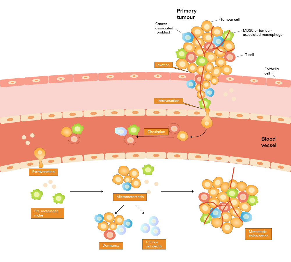
Recent studies have shown that the microenvironment of the target organ, often referred to as the "pre-metastatic niche," plays a significant role in determining where and how tumor cells will establish secondary growth. Factors such as inflammation, immune cell recruitment, and the remodeling of extracellular matrix (ECM) components create a favorable environment for metastatic cells to thrive. Understanding these processes is crucial for developing therapies that can interrupt the metastatic cascade at various stages, potentially inhibiting the spread of cancer before it becomes life-threatening.

Breast Cancer Metastasis

Detecting and managing breast cancer metastasis is particularly critical, as metastasis accounts for 90% of deaths in breast cancer patients despite advances in screening and treatment. Breast cancer metastasis typically involves the spread of malignant cells to bones, lungs, liver, and brain, leading to significant dysfunction in these organs. For instance, bone metastasis can result in severe pain and fractures, while brain metastasis may lead to neurological deficits and cognitive decline. The impact of such metastases on a patient's quality of life and survival cannot be overstated, making the detection and inhibition of breast cancer metastasis a major focus of ongoing research.

Immunohistochemical (IHC) analysis of specific biomarkers, such as HER2, Ki-67, and others, has proven instrumental in detecting breast cancer metastasis. Additionally, circulating tumor cells (CTCs) and cell-free DNA (cfDNA) in the bloodstream are emerging as non-invasive biomarkers that offer valuable insights into breast cancer's progression and potential spread. These techniques not only aid in early detection but also help in understanding the molecular mechanisms underlying breast cancer metastasis, which could lead to the development of targeted therapies.


Anti-HER2 Picoband Anti-Ki67 Picoband


Biomarker Detection

The detection of cancer metastasis is multifaceted and involves identifying various biological processes and signaling molecules associated with the metastatic cascade, including:

Epithelial-Mesenchymal Transition (EMT): During this process, epithelial cells in the primary tumor acquire mesenchymal properties, enhancing their mobility and invasiveness. Markers of EMT can serve as early indicators of metastatic potential.

Secretion of ECM Remodeling Enzymes: Cancer cells often secrete enzymes that degrade the extracellular matrix, facilitating invasion into surrounding tissues. Detection of these enzymes can provide insights into the aggressiveness of the tumor.

Angiogenesis Markers: Cancer cells invade tissues and stimulate the formation of new blood vessels (angiogenesis) to supply nutrients and oxygen. The presence of angiogenesis markers can indicate ongoing metastatic activity.

Quantification of CTCs: The number of CTCs in the blood correlates with the likelihood of metastasis and can be monitored to assess disease progression.

Colonization and Micro-Metastasis Formation: Monitoring the expression of specific genes and proteins involved in colonization can help predict the formation of micro-metastases, often precursors to larger metastatic tumors.


Boster Bio offers a comprehensive range of over 70 antibodies related to cancer metastasis and more than 200 breast cancer markers, providing researchers with the tools needed to investigate these critical processes. Here are some of them:

Inhibition

Inhibiting cancer metastasis is as crucial as detecting it, and strategies are often aligned with the steps of the metastatic process described above. Research has shown that inhibiting EMT, blocking ECM remodeling enzymes, and targeting angiogenesis can significantly reduce the spread of cancer. Antibodies against key proteins involved in these pathways, such as those targeting VEGF, MMPs, and EMT markers, are being developed and tested for their potential to inhibit metastasis.

In the context of breast cancer, targeting specific pathways involved in cell migration and invasion, such as the PI3K/AKT/mTOR pathway, has shown potential in preclinical studies. Additionally, novel biomarkers are being explored to identify high-risk patients who may benefit from more aggressive inhibition strategies. Combining these approaches with traditional therapies, such as chemotherapy and radiation, offers a more comprehensive method to inhibit metastasis and improve patient outcomes.


Anti-VEGF Picoband Anti-HER2 Picoband


External readings:

We have compiled some of the latest research on inhibiting breast cancer cell metastasis at the cellular level. Hope the unique strategies in these papers will inspire you.

Strategy 1: Activate the anticancer stimulator of interferon genes (STING)

The cGAMP is a cancer-cell-produced immunotransmitter that activates the STING pathway. And the ENPP1 is the dominant hydrolase of extracellular it. Studies have shown that reducing ENPP1 levels can decrease tumor growth and metastasis by enhancing the STING pathway, which activates an immune response. ENPP1 blockade represents a strategy to exploit cancer-produced extracellular cGAMP for controlled local activation of STING and is therefore a promising therapeutic approach against breast cancer.

Article name: ENPP1 is an innate immune checkpoint of the anticancer cGAMP-STING pathway in breast cancer

Authors: Wang Songnan, Böhnert Volker, Joseph Alby J, Sudaryo Valentino, et al.

Related ProductsGo to original article
cover 1

Strategy 2: Block EMT process

Epithelial-to-mesenchymal Transition (EMT) is a biological process in which epithelial cells lose their cell polarity and adhesion properties between cells. They become cells with the properties of mesenchymal cells. DAP5 promotes metastasis of breast cancer cells by affecting EMT. Silencing DAP5 significantly reduced cancer cell invasion, migration, metastasis, and survival in non-adherent conditions. Conversely, overexpression of DAP5 enhanced these processes.

Article name: Breast cancer cell mesenchymal transition and metastasis directed by DAP5/eIF3d-mediated selective mRNA translation

Authors: Amandine Alard, Olga Katsara, Tiffany Rios-Fuller, Columba de la Parra, et al.

Related ProductsGo to original website
cover2

Strategy 3: Prevent lysosomal degradation

Autophagy’s dysfunction is implicated in cancer and other diseases. Scientists employed an in vivo CRISPR screen targeting genes implicated in the regulation of autophagy to identify the Nsfl1c gene encoding p47 as a suppressor of human epidermal growth factor receptor 2 (HER2)+ breast cancer metastasis. Mechanistic studies show that p47 functions in the repair of lysosomal damage for autophagy flux and in the endosomal trafficking of nuclear factor κB, an essential modulator for lysosomal degradation to promote metastasis.

Article name: In vivo CRISPR knockout screen identifies p47 as a suppressor of HER2+ breast cancer metastasis by regulating NEMO trafficking and autophagy flux

Author: Amandine Alard, Olga Katsara, Tiffany Rios-Fuller, Columba de la Parra,et al.

Related ProductsGo to original website
cover3

Strategy 4: Change the microenvironment and use immune checkpoint blockade (ICB)


Triple-negative breast cancer is insensitive to ICB therapy due to insufficient CTL infiltration and a hyper-lactic acid-suppressive immune microenvironment from abnormal glycolysis. Scientists created CASN, a photodynamic bioregulator, from Chlorin e6 and MCT1 inhibitor AZD3965. CASN blocks MCT1-mediated lactic acid efflux, reducing lactic acid-induced regulatory T cell generation and M2 macrophage polarization, thereby attenuating immune suppression and promoting CTL recruitment and activation.

Article name: Carrier-Free Photodynamic Bioregulators Inhibiting Lactic Acid Efflux Combined with Immune Checkpoint Blockade for Triple-Negative Breast Cancer Immunotherapy

Authors:Guimei Chen, Ling Lin, Ziyi Mai , Yan Tang, et al.

Related ProductsGo to original website
cover4

More findings: New Biomarker circSIPA1L3

This study explores the role of circSIPA1L3 in breast cancer, highlighting its association with poor prognosis and its role in promoting tumor progression and glycolysis. The research shows that circSIPA1L3 is highly expressed in breast cancer tissues and serum, serving as a diagnostic and prognostic biomarker. Mechanistically, it stabilizes mRNA of glycolysis-related genes and enhances IGF2BP3-UPS7 interaction, promoting lactate secretion and tumor-associated macrophage recruitment. Elevated circSIPA1L3 levels correlate with unfavorable outcomes, suggesting it as a potential therapeutic target for breast cancer.

Article name: Exosomal circSIPA1L3-mediated intercellular communication contributes to glucose metabolic reprogramming and progression of triple-negative breast cancer

Author: Yiran Liang, Fangzhou Ye, Dan Luo, Li Long, &etc.

Related ProductsGo to original website
cover5