Click image to see more details
Product Info Summary
| SKU: | A15277-1 |
|---|---|
| Size: | 100 μg/vial |
| Reactive Species: | Human, Mouse, Rat |
| Host: | Rabbit |
| Application: | Flow Cytometry, IF, ICC, WB |
Customers Who Bought This Also Bought

Product info
Product Name
Anti-KBTBD2 Antibody Picoband®
SKU/Catalog Number
A15277-1
Size
100 μg/vial
Form
Lyophilized
Description
Boster Bio Anti-KBTBD2 Antibody Picoband® catalog # A15277-1. Tested in Flow Cytometry, IF, ICC, WB applications. This antibody reacts with Human, Mouse, Rat. The brand Picoband indicates this is a premium antibody that guarantees superior quality, high affinity, and strong signals with minimal background in Western blot applications. Only our best-performing antibodies are designated as Picoband, ensuring unmatched performance.
Storage & Handling
Store at -20˚C for one year from date of receipt. After reconstitution, at 4˚C for one month. It can also be aliquotted and stored frozen at -20˚C for six months. Avoid repeated freeze-thaw cycles.
Cite This Product
Anti-KBTBD2 Antibody Picoband® (Boster Biological Technology, Pleasanton CA, USA, Catalog # A15277-1)
Host
Rabbit
Contents
Each vial contains 4mg Trehalose, 0.9mg NaCl, 0.2mg Na2HPO4, 0.05mg NaN3.
Clonality
Polyclonal
Isotype
Rabbit IgG
Immunogen
A synthetic peptide corresponding to a sequence at the C-terminus of human KBTBD2, identical to the related mouse and rat sequences.
*Blocking peptide can be purchased. Costs vary based on immunogen length. Contact us for pricing.
Cross-reactivity
No cross-reactivity with other proteins.
Reactive Species
A15277-1 is reactive to KBTBD2 in Human, Mouse, Rat
Reconstitution
Add 0.2ml of distilled water will yield a concentration of 500ug/ml.
Observed Molecular Weight
72 kDa
Calculated molecular weight
24145 MW
Background of KBTBD2
This gene encodes a conserved protein that is similar to DNA-binding proteins, such as major centromere autoantigen B (CENPB). Inactivation of the related gene in mice resulted in epileptic seizures. Childhood Absence Epilepsy (CAE) has been mapped to the same chromosomal location (8q24.3) as this gene. Alternative splicing results in multiple transcript variants.
Antibody Validation
Boster validates all antibodies on WB, IHC, ICC, Immunofluorescence, and ELISA with known positive control and negative samples to ensure specificity and high affinity, including thorough antibody incubations.
Application & Images
Applications
A15277-1 is guaranteed for Flow Cytometry, IF, ICC, WB Boster Guarantee
Assay Dilutions Recommendation
The recommendations below provide a starting point for assay optimization. The actual working concentration varies and should be decided by the user.
Western blot, 0.1-0.5μg/ml
Immunocytochemistry/Immunofluorescence, 2μg/ml
Flow Cytometry (Fixed), 1-3μg/1x106 cells
Positive Control
WB: human Hela whole cell, human K562 whole cell, human U20S whole cell, human COLO320 whole cell, human SW620 whole cell, human Caco-2 whole cell, human placenta tissue, rat heart tissue, rat kidney tissue, rat PC-12 whole cell, mouse heart tissue, mouse kidney tissue
ICC/IF: A549 cell
FCM: U87 cell
Validation Images & Assay Conditions
Click image to see more details
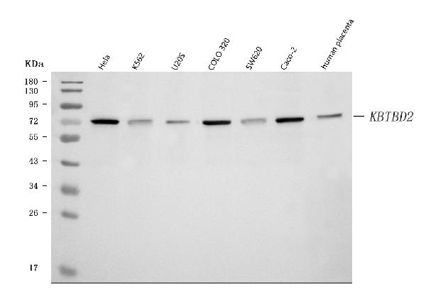
Figure 1. Western blot analysis of KBTBD2 using anti-KBTBD2 antibody (A15277-1).
Electrophoresis was performed on a 5-20% SDS-PAGE gel at 70V (Stacking gel) / 90V (Resolving gel) for 2-3 hours. The sample well of each lane was loaded with 30 ug of sample under reducing conditions.
Lane 1: human Hela whole cell lysates,
Lane 2: human K562 whole cell lysates,
Lane 3: human U20S whole cell lysates,
Lane 4: human COLO320 whole cell lysates,
Lane 5: human SW620 whole cell lysates,
Lane 6: human Caco-2 whole cell lysates,
Lane 7: human placenta tissue lysates.
After electrophoresis, proteins were transferred to a nitrocellulose membrane at 150 mA for 50-90 minutes. Blocked the membrane with 5% non-fat milk/TBS for 1.5 hour at RT. The membrane was incubated with rabbit anti-KBTBD2 antigen affinity purified polyclonal antibody (Catalog # A15277-1) at 0.5 μg/mL overnight at 4°C, then washed with TBS-0.1%Tween 3 times with 5 minutes each and probed with a goat anti-rabbit IgG-HRP secondary antibody at a dilution of 1:5000 for 1.5 hour at RT. The signal is developed using an Enhanced Chemiluminescent detection (ECL) kit (Catalog # EK1002) with Tanon 5200 system. A specific band was detected for KBTBD2 at approximately 72 kDa. The expected band size for KBTBD2 is at 71 kDa.

Click image to see more details
Figure 2. Western blot analysis of KBTBD2 using anti-KBTBD2 antibody (A15277-1).
Electrophoresis was performed on a 5-20% SDS-PAGE gel at 70V (Stacking gel) / 90V (Resolving gel) for 2-3 hours. The sample well of each lane was loaded with 30 ug of sample under reducing conditions.
Lane 1: rat heart tissue lysates,
Lane 2: rat kidney tissue lysates,
Lane 3: rat PC-12 whole cell lysates,
Lane 4: mouse heart tissue lysates,
Lane 5: mouse kidney tissue lysates.
After electrophoresis, proteins were transferred to a nitrocellulose membrane at 150 mA for 50-90 minutes. Blocked the membrane with 5% non-fat milk/TBS for 1.5 hour at RT. The membrane was incubated with rabbit anti-KBTBD2 antigen affinity purified polyclonal antibody (Catalog # A15277-1) at 0.5 μg/mL overnight at 4°C, then washed with TBS-0.1%Tween 3 times with 5 minutes each and probed with a goat anti-rabbit IgG-HRP secondary antibody at a dilution of 1:5000 for 1.5 hour at RT. The signal is developed using an Enhanced Chemiluminescent detection (ECL) kit (Catalog # EK1002) with Tanon 5200 system. A specific band was detected for KBTBD2 at approximately 72 kDa. The expected band size for KBTBD2 is at 71 kDa.

Click image to see more details
Figure 4. Flow Cytometry analysis of U87 cells using anti-KBTBD2 antibody (A15277-1).
Overlay histogram showing U87 cells stained with A15277-1 (Blue line). To facilitate intracellular staining, cells were fixed with 4% paraformaldehyde and permeabilized with permeabilization buffer. The cells were blocked with 10% normal goat serum. And then incubated with rabbit anti-KBTBD2 Antibody (A15277-1, 1μg/1x106 cells) for 30 min at 20°C. DyLight®488 conjugated goat anti-rabbit IgG (BA1127, 5-10μg/1x106 cells) was used as secondary antibody for 30 minutes at 20°C. Isotype control antibody (Green line) was rabbit IgG (1μg/1x106) used under the same conditions. Unlabelled sample without incubation with primary antibody and secondary antibody (Red line) was used as a blank control.

Click image to see more details
Figure 3. IF analysis of KBTBD2 using anti-KBTBD2 antibody (A15277-1).
KBTBD2 was detected in immunocytochemical section of A549 cell. Enzyme antigen retrieval was performed using IHC enzyme antigen retrieval reagent (AR0022) for 15 mins. The cells were blocked with 10% goat serum. And then incubated with 2μg/mL rabbit anti-KBTBD2 Antibody (A15277-1) overnight at 4°C. DyLight®488 Conjugated Goat Anti-Rabbit IgG (BA1127) was used as secondary antibody at 1:100 dilution and incubated for 30 minutes at 37°C. The section was counterstained with DAPI. Visualize using a fluorescence microscope and filter sets appropriate for the label used.
Protein Target Info & Infographic
Gene/Protein Information For KBTBD2 (Source: Uniprot.org, NCBI)
Gene Name
KBTBD2
Full Name
Kelch repeat and BTB domain-containing protein 2
Weight
24145 MW
Alternative Names
Kelch repeat and BTB domain-containing protein 2; BTB and kelch domain-containing protein 1; KBTBD2; BKLHD1; KIAA1489 KBTBD2 BKLHD1 kelch repeat and BTB domain containing 2 kelch repeat and BTB domain-containing protein 2|BTB and kelch domain containing 1|BTB and kelch domain-containing protein 1|kelch repeat and BTB (POZ) domain containing 2
*If product is indicated to react with multiple species, protein info is based on the gene entry specified above in "Species".For more info on KBTBD2, check out the KBTBD2 Infographic

We have 30,000+ of these available, one for each gene! Check them out.
In this infographic, you will see the following information for KBTBD2: database IDs, superfamily, protein function, synonyms, molecular weight, chromosomal locations, tissues of expression, subcellular locations, post-translational modifications, and related diseases, research areas & pathways. If you want to see more information included, or would like to contribute to it and be acknowledged, please contact [email protected].
Specific Publications For Anti-KBTBD2 Antibody Picoband® (A15277-1)
Loading publications
Recommended Resources
Here are featured tools and databases that you might find useful.
- Boster's Pathways Library
- Protein Databases
- Bioscience Research Protocol Resources
- Data Processing & Analysis Software
- Photo Editing Software
- Scientific Literature Resources
- Research Paper Management Tools
- Molecular Biology Software
- Primer Design Tools
- Bioinformatics Tools
- Phylogenetic Tree Analysis
Customer Reviews
Have you used Anti-KBTBD2 Antibody Picoband®?
Submit a review and receive an Amazon gift card.
- $30 for a review with an image
0 Reviews For Anti-KBTBD2 Antibody Picoband®
Customer Q&As
Have a question?
Find answers in Q&As, reviews.
Can't find your answer?
Submit your question
3 Customer Q&As for Anti-KBTBD2 Antibody Picoband®
Question
We are currently using anti-KBTBD2 antibody A15277-1 for human tissue, and we are well pleased with the WB results. The species of reactivity given in the datasheet says human, mouse, rat. Is it possible that the antibody can work on feline tissues as well?
Verified Customer
Verified customer
Asked: 2020-03-24
Answer
The anti-KBTBD2 antibody (A15277-1) has not been tested for cross reactivity specifically with feline tissues, but there is a good chance of cross reactivity. We have an innovator award program that if you test this antibody and show it works in feline you can get your next antibody for free. Please contact me if I can help you with anything.
Boster Scientific Support
Answered: 2020-03-24
Question
I was wanting to use your anti-KBTBD2 antibody for WB for mouse brain on frozen tissues, but I want to know if it has been tested for this particular application. Has this antibody been tested and is this antibody a good choice for mouse brain identification?
R. Bhatt
Verified customer
Asked: 2019-09-13
Answer
As indicated on the product datasheet, A15277-1 anti-KBTBD2 antibody has been validated for IF, ICC, WB on human, mouse, rat tissues. We have an innovator award program that if you test this antibody and show it works in mouse brain in IHC-frozen, you can get your next antibody for free.
Boster Scientific Support
Answered: 2019-09-13
Question
I have a question about product A15277-1, anti-KBTBD2 antibody. I was wondering if it would be possible to conjugate this antibody with biotin. I would need it to be without BSA or sodium azide. I am planning on using a buffer exchange of sodium azide with PBS only. Would there be problems for me to conjugate the antibody and store it in -20 degrees in small aliquots?
Verified Customer
Verified customer
Asked: 2018-06-27
Answer
We do not advise storing this antibody with PBS buffer only in -20 degrees. If you want to store it in -20 degrees it is best to add some cryoprotectant like glycerol. If you want carrier free A15277-1 anti-KBTBD2 antibody, we can provide it to you in a special formula with trehalose and/or glycerol. These molecules will not interfere with conjugation chemistry and provide a good level of protection for the antibody from degradation. Please be sure to specify this in your purchase order.
Boster Scientific Support
Answered: 2018-06-27
