This website uses cookies to ensure you get the best experience on our website.
- Table of Contents
1 Citations 16 Q&As
8 Citations 17 Q&As


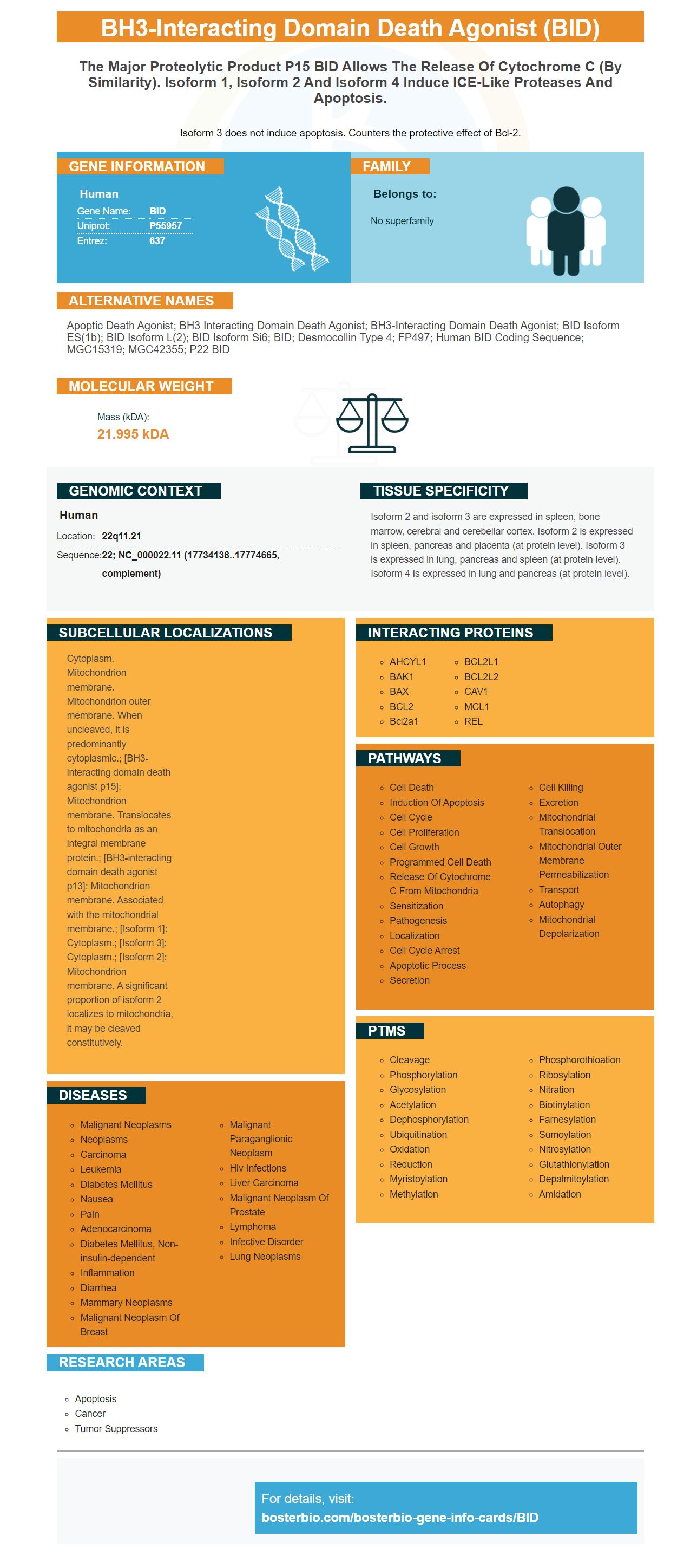
Facts about BH3-interacting domain death agonist.

Isoform 3 does not induce apoptosis. Counters the protective effect of Bcl-2.
| Human | |
|---|---|
| Gene Name: | BID |
| Uniprot: | P55957 |
| Entrez: | 637 |

| Belongs to: |
|---|
| No superfamily |

apoptic death agonist; BH3 interacting domain death agonist; BH3-interacting domain death agonist; BID isoform ES(1b); BID isoform L(2); BID isoform Si6; BID; desmocollin type 4; FP497; Human BID coding sequence; MGC15319; MGC42355; p22 BID
Mass (kDA):
21.995 kDA

| Human | |
|---|---|
| Location: | 22q11.21 |
| Sequence: | 22; NC_000022.11 (17734138..17774665, complement) |
Isoform 2 and isoform 3 are expressed in spleen, bone marrow, cerebral and cerebellar cortex. Isoform 2 is expressed in spleen, pancreas and placenta (at protein level). Isoform 3 is expressed in lung, pancreas and spleen (at protein level). Isoform 4 is expressed in lung and pancreas (at protein level).
Cytoplasm. Mitochondrion membrane. Mitochondrion outer membrane. When uncleaved, it is predominantly cytoplasmic.; [BH3-interacting domain death agonist p15]: Mitochondrion membrane. Translocates to mitochondria as an integral membrane protein.; [BH3-interacting domain death agonist p13]: Mitochondrion membrane. Associated with the mitochondrial membrane.; [Isoform 1]: Cytoplasm.; [Isoform 3]: Cytoplasm.; [Isoform 2]: Mitochondrion membrane. A significant proportion of isoform 2 localizes to mitochondria, it may be cleaved constitutively.


PMID: 8918887 by Wang K., et al. BID: a novel BH3 domain-only death agonist.
PMID: 9721221 by Footz T.K., et al. The gene for death agonist BID maps to the region of human 22q11.2 duplicated in cat eye syndrome chromosomes and to mouse chromosome 6.
*More publications can be found for each product on its corresponding product page