This website uses cookies to ensure you get the best experience on our website.
- Table of Contents
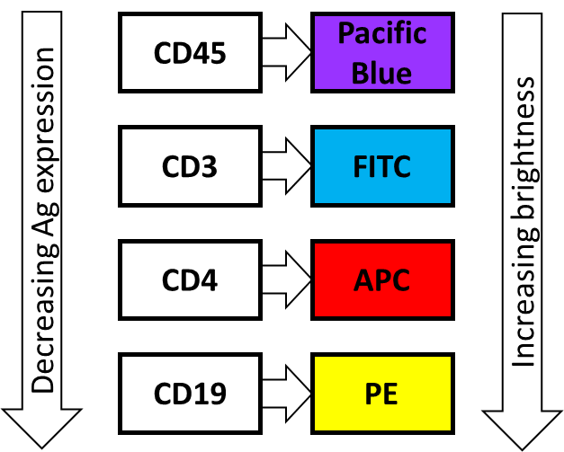
Always try to pair the brightest fluorochromes with the weakest expressing antigen. In case the expression level of an antigen is unknown, it is advisable to use brighter fluorochromes.
Avoid spillover or spectral overlap between fluorochromes by spreading them as much as possible across the spectrum.
Avoid fluorochromes that can be excited by more than one laser e.g. APC-Cy7.
Always include suitable compensation controls especially if points 3 and 4 above cannot be achieved.
It is also advisable to use FMO controls to gate differently stained sub-populations more accurately.
Consider using online multicolor panel designers provided by FACS technology companies.
A suitable 4-color panel to illustrate points 1-3. The color of the boxes correspond to the lasers that excite the respective fluorochromes.

Keywords: FACS fluorophores, fluorochrome conjugates, multi color panel, flow validated antibody panel, excitation emission, FACS dyes
Click for more optimization tipsProtocols, optimization tips, troubleshooting guides, and more for flow cytometry.
Technical ResourcesDownload troubleshooting handbooks for IHC, Western blot and ELISA for FREE.
Troubleshooting GuidesGet some of the best Bosterbio's flow cytometry optimization tips. This guide includes protocols, optimization tips, troubleshooting tips, and more on flow cytometry.
See MoreBoster Bio is an antibody company and supplier. Read more about our troubleshooting tips for flow cytometry (FACS) experiments. Learn tips on how to resolve issues such as high background, and weak or no signal
See MoreFlow cytometry is a popular cell biology technique that utilizes laser-based technology to count, sort, and profile cells in a heterogeneous fluid mixture. FACS is an abbreviation for fluorescence-activated cell sorting, which is a flow cytometry technique that further adds a degree of functionality.
See MoreAntibody titration is recommended to determine the correct concentration of an antibody for the optimum signal. Test different dilutions of the antibody to zero in on the lowest concentration that gives the strongest signal in positive control and th...
See MoreThe principle behind Immunohistochemistry (IHC) entails detection of antigen or happens in cells of a tissue section by exploiting the principle of antibodies binding specifically to antigens in biological tissues. Learn more about this in this guide.
See MoreThe choice of which detection system to use is influenced by the equipment available and the needs of the specific application being considered. Use this guide to help decide whether to use enzyme-linked secondary antibodies, or fluorescently labelled secondary antibodies as your detection system.
See MoreThis guide provides a thorough list of Immunohistochemistry troubleshooting tips, including weak staining, high background, nonspecific staining among others. Learn how to take control of your IHC process.
See MoreUse this guide to help decide whether to use enzyme-linked secondary antibodies, or fluorescently labelled secondary antibodies as your detection system. ELISA Blocking Buffer Optimization There are a variety of blocking buffers, not one of which is ...
See More