Click image to see more details
Product Info Summary
| SKU: | A00210-1 |
|---|---|
| Size: | 100 μg/vial |
| Reactive Species: | Human, Mouse, Rat |
| Host: | Rabbit |
| Application: | Flow Cytometry, WB |
Customers Who Bought This Also Bought


Product info
Product Name
Anti-Vitamin D Receptor/VDR Antibody Picoband®
View all VDR/NR1I1/Vitamin D Receptor Antibodies
SKU/Catalog Number
A00210-1
Size
100 μg/vial
Form
Lyophilized
Description
Boster Bio Anti-Vitamin D Receptor/VDR Antibody Picoband® catalog # A00210-1. Tested in Flow Cytometry, WB applications. This antibody reacts with Human, Mouse, Rat. The brand Picoband indicates this is a premium antibody that guarantees superior quality, high affinity, and strong signals with minimal background in Western blot applications. Only our best-performing antibodies are designated as Picoband, ensuring unmatched performance.
Storage & Handling
At -20°C for one year from date of receipt. After reconstitution, at 4°C for one month. It can also be aliquotted and stored frozen at -20°C for six months. Avoid repeated freezing and thawing.
Cite This Product
Anti-Vitamin D Receptor/VDR Antibody Picoband® (Boster Biological Technology, Pleasanton CA, USA, Catalog # A00210-1)
Host
Rabbit
Contents
Each vial contains 4 mg Trehalose, 0.9 mg NaCl, 0.2 mg Na2HPO4.
Clonality
Polyclonal
Isotype
Rabbit IgG
Immunogen
A synthetic peptide corresponding to a sequence at the C-terminus of human Vitamin D Receptor/VDR, which shares 88.9% amino acid (aa) sequence identity with mouse and rat VDR.
*Blocking peptide can be purchased. Costs vary based on immunogen length. Contact us for pricing.
Cross-reactivity
No cross-reactivity with other proteins.
Reactive Species
A00210-1 is reactive to VDR in Human, Mouse, Rat
Reconstitution
Adding 0.2 ml of distilled water will yield a concentration of 500 μg/ml.
Observed Molecular Weight
60 kDa
Calculated molecular weight
24423 MW
Background of VDR/NR1I1/Vitamin D Receptor
VDR (Vitamin D Receptor), also known as Vitamin D Hormone Receptor, is a member of the nuclear receptor family of transcription factors. Labuda et al. (1991) assigned the VDR gene to 12q12-q14 by in situ hybridization. Using mutation analysis, Jurutka et al. (2000) characterized arg18/arg22, VDR residues immediately N-terminal of the first DNA-binding zinc finger, as vital for contact with the general transcription factor IIB (TFIIB). A natural polymorphic variant of VDR, termed F/M4 (missing a FokI restriction site), which lacks only the first 3 amino acids (including glu2), interacted more efficiently with TFIIB and also possessed elevated transcriptional activity compared with the full-length (f/M1) receptor. Shah et al. (2006) stated that the signaling and oncogenic activity of beta-catenin (CTNNB1) can be repressed by activation of VDR. Conversely, high levels of beta-catenin can potentiate the transcriptional activity of 1,25- dihydroxyvitamin D3.
Antibody Validation
Boster validates all antibodies on WB, IHC, ICC, Immunofluorescence, and ELISA with known positive control and negative samples to ensure specificity and high affinity, including thorough antibody incubations.
Application & Images
Applications
A00210-1 is guaranteed for Flow Cytometry, WB Boster Guarantee
Assay Dilutions Recommendation
The recommendations below provide a starting point for assay optimization. The actual working concentration varies and should be decided by the user.
Western blot, 0.25-0.5 μg/ml, Human, Mouse, Rat
Flow Cytometry (Fixed), 1-3 μg/1x106 cells, Human, Rat
Positive Control
WB: human SH-SY5Y whole cell, human U-937 whole cell, human Hacat whole cell, human HL-60 whole cell, rat brain tissue, rat PC-12 whole cell, mouse brain tissue, mouse NIH/3T3 whole cell
FCM: C6 cell, U20S cell
Validation Images & Assay Conditions
Click image to see more details
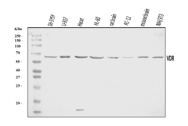
Figure 1. Western blot analysis of Vitamin D Receptor/VDR using anti-Vitamin D Receptor/VDR antibody (A00210-1).
Electrophoresis was performed on a 5-20% SDS-PAGE gel at 70V (Stacking gel) / 90V (Resolving gel) for 2-3 hours. The sample well of each lane was loaded with 30 ug of sample under reducing conditions.
Lane 1: human SH-SY5Y whole cell lysates,
Lane 2: human U-937 whole cell lysates,
Lane 3: human Hacat whole cell lysates,
Lane 4: human HL-60 whole cell lysates,
Lane 5: rat brain tissue lysates,
Lane 6: rat PC-12 whole cell lysates,
Lane 7: mouse brain tissue lysates,
Lane 8: mouse NIH/3T3 whole cell lysates.
After electrophoresis, proteins were transferred to a nitrocellulose membrane at 150 mA for 50-90 minutes. Blocked the membrane with 5% non-fat milk/TBS for 1.5 hour at RT. The membrane was incubated with rabbit anti-Vitamin D Receptor/VDR antigen affinity purified polyclonal antibody (Catalog # A00210-1) at 0.5 μg/mL overnight at 4°C, then washed with TBS-0.1%Tween 3 times with 5 minutes each and probed with a goat anti-rabbit IgG-HRP secondary antibody at a dilution of 1:5000 for 1.5 hour at RT. The signal is developed using an Enhanced Chemiluminescent detection (ECL) kit (Catalog # EK1002) with Tanon 5200 system. A specific band was detected for Vitamin D Receptor/VDR at approximately 60 kDa. The expected band size for Vitamin D Receptor/VDR is at 60 kDa.

Click image to see more details
Figure 2. Flow Cytometry analysis of C6 cells using anti-Vitamin D Receptor/VDR antibody (A00210-1).
Overlay histogram showing C6 cells stained with A00210-1 (Blue line). To facilitate intracellular staining, cells were fixed with 4% paraformaldehyde and permeabilized with permeabilization buffer. The cells were blocked with 10% normal goat serum. And then incubated with rabbit anti-Vitamin D Receptor/VDR Antibody (A00210-1, 1 μg/1x106 cells) for 30 min at 20°C. DyLight®488 conjugated goat anti-rabbit IgG (BA1127, 5-10 μg/1x106 cells) was used as secondary antibody for 30 minutes at 20°C. Isotype control antibody (Green line) was rabbit IgG (1 μg/1x106) used under the same conditions. Unlabelled sample without incubation with primary antibody and secondary antibody (Red line) was used as a blank control.

Click image to see more details
Figure 3. Flow Cytometry analysis of U20S cells using anti-Vitamin D Receptor/VDR antibody (A00210-1).
Overlay histogram showing U20S cells stained with A00210-1 (Blue line). To facilitate intracellular staining, cells were fixed with 4% paraformaldehyde and permeabilized with permeabilization buffer. The cells were blocked with 10% normal goat serum. And then incubated with rabbit anti-Vitamin D Receptor/VDR Antibody (A00210-1, 1 μg/1x106 cells) for 30 min at 20°C. DyLight®488 conjugated goat anti-rabbit IgG (BA1127, 5-10 μg/1x106 cells) was used as secondary antibody for 30 minutes at 20°C. Isotype control antibody (Green line) was rabbit IgG (1 μg/1x106) used under the same conditions. Unlabelled sample without incubation with primary antibody and secondary antibody (Red line) was used as a blank control.
Protein Target Info & Infographic
Gene/Protein Information For VDR (Source: Uniprot.org, NCBI)
Gene Name
VDR
Full Name
Vitamin D3 receptor
Weight
24423 MW
Superfamily
nuclear hormone receptor family
Alternative Names
GTP-binding nuclear protein Ran;Androgen receptor-associated protein 24;GTPase Ran;Ras-like protein TC4;Ras-related nuclear protein;RAN;ARA24;OK/SW-cl.81; VDR NR1I1, PPP1R163 vitamin D receptor vitamin D3 receptor|1,25-dihydroxyvitamin D3 receptor|nuclear receptor subfamily 1 group I member 1|protein phosphatase 1, regulatory subunit 163|vitamin D (1,25- dihydroxyvitamin D3) receptor|vitamin D nuclear receptor variant 1
*If product is indicated to react with multiple species, protein info is based on the gene entry specified above in "Species".For more info on VDR, check out the VDR Infographic

We have 30,000+ of these available, one for each gene! Check them out.
In this infographic, you will see the following information for VDR: database IDs, superfamily, protein function, synonyms, molecular weight, chromosomal locations, tissues of expression, subcellular locations, post-translational modifications, and related diseases, research areas & pathways. If you want to see more information included, or would like to contribute to it and be acknowledged, please contact [email protected].
Specific Publications For Anti-Vitamin D Receptor/VDR Antibody Picoband® (A00210-1)
Hello CJ!
No publications found for A00210-1
*Do you have publications using this product? Share with us and receive a reward. Ask us for more details.
Recommended Resources
Here are featured tools and databases that you might find useful.
- Boster's Pathways Library
- Protein Databases
- Bioscience Research Protocol Resources
- Data Processing & Analysis Software
- Photo Editing Software
- Scientific Literature Resources
- Research Paper Management Tools
- Molecular Biology Software
- Primer Design Tools
- Bioinformatics Tools
- Phylogenetic Tree Analysis
Customer Reviews
Have you used Anti-Vitamin D Receptor/VDR Antibody Picoband®?
Submit a review and receive an Amazon gift card.
- $30 for a review with an image
0 Reviews For Anti-Vitamin D Receptor/VDR Antibody Picoband®
Customer Q&As
Have a question?
Find answers in Q&As, reviews.
Can't find your answer?
Submit your question
1 Customer Q&As for Anti-Vitamin D Receptor/VDR Antibody Picoband®
Question
Could you please tell me the composition buffer before lyophilisation? How much sodium azide is in this product?
Verified customer
Asked: 2022-09-30
Answer
The composition buffer before lyophilisation for A00210-1 is 40 mg /ml Trehalose, 9mg /mlNaCl and 2mg /mlNa2HPO4. This product doesn't contain any sodium azide.
Boster Scientific Support
Answered: 2022-09-30
