Click image to see more details
Product Info Summary
| SKU: | A06313-2 |
|---|---|
| Size: | 100 μg/vial |
| Reactive Species: | Human, Mouse, Rat |
| Host: | Rabbit |
| Application: | ELISA, WB |
Customers Who Bought This Also Bought

Product info
Product Name
Anti-TUBG1/2 Antibody Picoband®
SKU/Catalog Number
A06313-2
Size
100 μg/vial
Form
Lyophilized
Description
Boster Bio Anti-TUBG1/2 Antibody Picoband® catalog # A06313-2. Tested in ELISA, WB applications. This antibody reacts with Human, Mouse, Rat. The brand Picoband indicates this is a premium antibody that guarantees superior quality, high affinity, and strong signals with minimal background in Western blot applications. Only our best-performing antibodies are designated as Picoband, ensuring unmatched performance.
Storage & Handling
At -20°C for one year from date of receipt. After reconstitution, at 4°C for one month. It can also be aliquotted and stored frozen at -20°C for six months. Avoid repeated freezing and thawing.
Cite This Product
Anti-TUBG1/2 Antibody Picoband® (Boster Biological Technology, Pleasanton CA, USA, Catalog # A06313-2)
Host
Rabbit
Contents
Each vial contains 4 mg Trehalose, 0.9 mg NaCl, 0.2 mg Na2HPO4.
Clonality
Polyclonal
Isotype
Rabbit IgG
Immunogen
E.coli-derived human TUBG1/2 recombinant protein (Position: Q167-Q394).
*Blocking peptide can be purchased. Costs vary based on immunogen length. Contact us for pricing.
Cross-reactivity
No cross-reactivity with other proteins.
Reactive Species
A06313-2 is reactive to TUBG1/2 in Human, Mouse, Rat
Reconstitution
Adding 0.2 ml of distilled water will yield a concentration of 500 µg/ml.
Observed Molecular Weight
51 kDa
Calculated molecular weight
49655 MW
Background of TUBG1/2
Tubulin, gamma 1/2 is a protein in humans that is encoded by the TUBG1/2 gene. This gene encodes a member of the tubulin superfamily. The encoded protein localizes to the centrosome where it binds to microtubules as part of a complex referred to as the gamma-tubulin ring complex. The protein mediates microtubule nucleation and is required for microtubule formation and progression of the cell cycle. A pseudogene of this gene is found on chromosome 7.
Antibody Validation
Boster validates all antibodies on WB, IHC, ICC, Immunofluorescence, and ELISA with known positive control and negative samples to ensure specificity and high affinity, including thorough antibody incubations.
Application & Images
Applications
A06313-2 is guaranteed for ELISA, WB Boster Guarantee
Assay Dilutions Recommendation
The recommendations below provide a starting point for assay optimization. The actual working concentration varies and should be decided by the user.
Western blot, 0.1-0.25 µg/ml, Human, Mouse, Rat
ELISA, 0.1-0.5 µg/ml, Human
Positive Control
WB: human MCF-7 whole cell, human HepG2 whole cell, human A549 whole cell, human T-47D whole cell, human U251 whole cell, human Caco-2 whole cell, human Hacat whole cell, rat testis tissue, rat brain tissue, rat C6 whole cell, rat PC-12 whole cell, mouse testis tissue, mouse brain tissue, mouse Neuro-2a whole cell, mouse SP2/0 whole cell
Validation Images & Assay Conditions
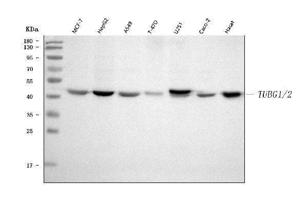
Click image to see more details
Figure 1. Western blot analysis of TUBG1/2 using anti-TUBG1/2 antibody (A06313-2).
Electrophoresis was performed on a 5-20% SDS-PAGE gel at 70V (Stacking gel) / 90V (Resolving gel) for 2-3 hours. The sample well of each lane was loaded with 30 ug of sample unde
r reducing conditions.
Lane 1: human MCF-7 whole cell lysates,
Lane 2: human HepG2 whole cell lysates,
Lane 3: human A549 whole cell lysates,
Lane 4: human T-47D whole cell lysates,
Lane 5: human U251 whole cell lysates,
Lane 6: human Caco-2 whole cell lysates,
Lane 7: human Hacat whole cell lysates.
After electrophoresis, proteins were transferred to a nitrocellulose membrane at 150 mA for 50-90 minutes. Blocked the membrane with 5% non-fat milk/TBS for 1.5 hour at RT. The membrane was incubated with rabbit anti-TUBG1/2 antigen affinity purified polyclonal antibody (Catalog # A06313-2) at 0.25 μg/mL overnight at 4°C, then washed with TBS-0.1%Tween 3 times with 5 minutes each and probed with a goat anti-rabbit IgG-HRP secondary antibody at a dilution of 1:5000 for 1.5 hour at RT. The signal is developed using an Enhanced Chemiluminescent detection (ECL) kit (Catalog # EK1002) with Tanon 5200 system. A specific band was detected for TUBG1/2 at approximately 51 kDa. The expected band size for TUBG1/2 is at 51 kDa.

Click image to see more details
Figure 2. Western blot analysis of TUBG1/2 using anti-TUBG1/2 antibody (A06313-2).
Electrophoresis was performed on a 5-20% SDS-PAGE gel at 70V (Stacking gel) / 90V (Resolving gel) for 2-3 hours. The sample well of each lane was loaded with 30 ug of sample unde
r reducing conditions.
Lane 1: rat testis tissue lysates,
Lane 2: rat brain tissue lysates,
Lane 3: rat C6 whole cell lysates,
Lane 4: rat PC-12 whole cell lysates,
Lane 5: mouse testis tissue lysates,
Lane 6: mouse brain tissue lysates,
Lane 7: mouse Neuro-2a whole cell lysates,
Lane 8: mouse SP2/0 whole cell lysates.
After electrophoresis, proteins were transferred to a nitrocellulose membrane at 150 mA for 50-90 minutes. Blocked the membrane with 5% non-fat milk/TBS for 1.5 hour at RT. The membrane was incubated with rabbit anti-TUBG1/2 antigen affinity purified polyclonal antibody (Catalog # A06313-2) at 0.25 μg/mL overnight at 4°C, then washed with TBS-0.1%Tween 3 times with 5 minutes each and probed with a goat anti-rabbit IgG-HRP secondary antibody at a dilution of 1:5000 for 1.5 hour at RT. The signal is developed using an Enhanced Chemiluminescent detection (ECL) kit (Catalog # EK1002) with Tanon 5200 system. A specific band was detected for TUBG1/2 at approximately 51 kDa. The expected band size for TUBG1/2 is at 51 kDa.
Protein Target Info & Infographic
Gene/Protein Information For TUBG1/2 (Source: Uniprot.org, NCBI)
Gene Name
TUBG1/2
Full Name
Weight
49655 MW
Alternative Names
ELAV-like protein 2; ELAV-like neuronal protein 1; Hu-antigen B; HuB; Nervous system-specific RNA-binding protein Hel-N1; ELAVL2; HUB
*If product is indicated to react with multiple species, protein info is based on the gene entry specified above in "Species".For more info on TUBG1/2, check out the TUBG1/2 Infographic

We have 30,000+ of these available, one for each gene! Check them out.
In this infographic, you will see the following information for TUBG1/2: database IDs, superfamily, protein function, synonyms, molecular weight, chromosomal locations, tissues of expression, subcellular locations, post-translational modifications, and related diseases, research areas & pathways. If you want to see more information included, or would like to contribute to it and be acknowledged, please contact [email protected].
Specific Publications For Anti-TUBG1/2 Antibody Picoband® (A06313-2)
Hello CJ!
No publications found for A06313-2
*Do you have publications using this product? Share with us and receive a reward. Ask us for more details.
Recommended Resources
Here are featured tools and databases that you might find useful.
- Boster's Pathways Library
- Protein Databases
- Bioscience Research Protocol Resources
- Data Processing & Analysis Software
- Photo Editing Software
- Scientific Literature Resources
- Research Paper Management Tools
- Molecular Biology Software
- Primer Design Tools
- Bioinformatics Tools
- Phylogenetic Tree Analysis
Customer Reviews
Have you used Anti-TUBG1/2 Antibody Picoband®?
Submit a review and receive an Amazon gift card.
- $30 for a review with an image
0 Reviews For Anti-TUBG1/2 Antibody Picoband®
Customer Q&As
Have a question?
Find answers in Q&As, reviews.
Can't find your answer?
Submit your question
