Click image to see more details
Product Info Summary
| SKU: | A02151-3 |
|---|---|
| Size: | 100 μg/vial |
| Reactive Species: | Human, Mouse, Rat |
| Host: | Rabbit |
| Application: | ELISA, Flow Cytometry, WB |
Customers Who Bought This Also Bought


Product info
Product Name
Anti-TAF1 Antibody Picoband®
View all KAT4/TBP Associated Factor 1 Antibodies
SKU/Catalog Number
A02151-3
Size
100 μg/vial
Form
Lyophilized
Description
Boster Bio Anti-TAF1 Antibody Picoband® catalog # A02151-3. Tested in ELISA, Flow Cytometry, WB applications. This antibody reacts with Human, Mouse, Rat. The brand Picoband indicates this is a premium antibody that guarantees superior quality, high affinity, and strong signals with minimal background in Western blot applications. Only our best-performing antibodies are designated as Picoband, ensuring unmatched performance.
Storage & Handling
At -20°C for one year from date of receipt. After reconstitution, at 4°C for one month. It can also be aliquotted and stored frozen at -20°C for six months. Avoid repeated freezing and thawing.
Cite This Product
Anti-TAF1 Antibody Picoband® (Boster Biological Technology, Pleasanton CA, USA, Catalog # A02151-3)
Host
Rabbit
Contents
Each vial contains 4 mg Trehalose, 0.9 mg NaCl, 0.2 mg Na2HPO4.
Clonality
Polyclonal
Isotype
Rabbit IgG
Immunogen
E.coli-derived human TAF1 recombinant protein (Position: H1375-D1636).
*Blocking peptide can be purchased. Costs vary based on immunogen length. Contact us for pricing.
Cross-reactivity
No cross-reactivity with other proteins.
Reactive Species
A02151-3 is reactive to TAF1 in Human, Mouse, Rat
Reconstitution
Adding 0.2 ml of distilled water will yield a concentration of 500 μg/ml.
Observed Molecular Weight
250 kDa
Calculated molecular weight
212.677kDa
Background of KAT4/TBP Associated Factor 1
Transcription initiation factor TFIID subunit 1, also known as transcription initiation factor TFIID 250 kDa subunit (TAFII-250) or TBP-associated factor 250 kDa (p250), is a protein that in humans is encoded by the TAF1 gene. Initiation of transcription by RNA polymerase II requires the activities of more than 70 polypeptides. The protein that coordinates these activities is the basal transcription factor TFIID, which binds to the core promoter to position the polymerase properly, serves as the scaffold for assembly of the remainder of the transcription complex, and acts as a channel for regulatory signals. TFIID is composed of the TATA-binding protein (TBP) and a group of evolutionarily conserved proteins known as TBP-associated factors or TAFs. TAFs may participate in basal transcription, serve as coactivators, function in promoter recognition or modify general transcription factors (GTFs) to facilitate complex assembly and transcription initiation. This gene encodes the largest subunit of TFIID. This subunit binds to core promoter sequences encompassing the transcription start site. It also binds to activators and other transcriptional regulators, and these interactions affect the rate of transcription initiation. This subunit contains two independent protein kinase domains at the N- and C-terminals, but also possesses acetyltransferase activity and can act as a ubiquitin-activating/conjugating enzyme. Mutations in this gene result in Dystonia 3, torsion, X-linked, a dystonia-parkinsonism disorder. Alternative splicing of this gene results in multiple transcript variants. This gene is part of a complex transcription unit (TAF1/DYT3), wherein some transcript variants share exons with TAF1 as well as additional downstream DYT3 exons.
Antibody Validation
Boster validates all antibodies on WB, IHC, ICC, Immunofluorescence, and ELISA with known positive control and negative samples to ensure specificity and high affinity, including thorough antibody incubations.
Application & Images
Applications
A02151-3 is guaranteed for ELISA, Flow Cytometry, WB Boster Guarantee
Assay Dilutions Recommendation
The recommendations below provide a starting point for assay optimization. The actual working concentration varies and should be decided by the user.
Western blot, 0.25-0.5 μg/ml, Human, Mouse, Rat
Flow Cytometry (Fixed), 1-3 μg/1x106 cells, Human
ELISA, 0.1-0.5 μg/ml, Human
Positive Control
WB: human Hela whole cell, human HepG2 whole cell, human 293T whole cell, human Jurkat whole cell, rat bain tissue, mouse brain tissue, mouse lung tissue
FCM: THP-1 cell
Validation Images & Assay Conditions
Click image to see more details
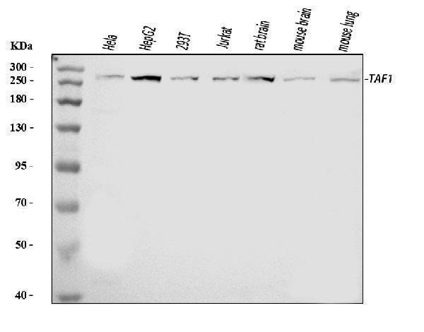
Figure 1. Western blot analysis of TAF1 using anti-TAF1 antibody (A02151-3).
Electrophoresis was performed on a 5-20% SDS-PAGE gel at 70V (Stacking gel) / 90V (Resolving gel) for 2-3 hours. The sample well of each lane was loaded with 30 ug of sample under reducing conditions.
Lane 1: human Hela whole cell lysates,
Lane 2: human HepG2 whole cell lysates,
Lane 3: human 293T whole cell lysates,
Lane 4: human Jurkat whole cell lysates,
Lane 5: rat bain tissue lysates,
Lane 6: mouse brain tissue lysates,
Lane 7: mouse lung tissue lysates.
After electrophoresis, proteins were transferred to a nitrocellulose membrane at 150 mA for 50-90 minutes. Blocked the membrane with 5% non-fat milk/TBS for 1.5 hour at RT. The membrane was incubated with rabbit anti-TAF1 antigen affinity purified polyclonal antibody (Catalog # A02151-3) at 0.5 μg/mL overnight at 4°C, then washed with TBS-0.1%Tween 3 times with 5 minutes each and probed with a goat anti-rabbit IgG-HRP secondary antibody at a dilution of 1:5000 for 1.5 hour at RT. The signal is developed using an Enhanced Chemiluminescent detection (ECL) kit (Catalog # EK1002) with Tanon 5200 system. A specific band was detected for TAF1 at approximately 250 kDa. The expected band size for TAF1 is at 250 kDa.

Click image to see more details
Figure 2. Flow Cytometry analysis of THP-1 cells using anti-TAF1 antibody (A02151-3).
Overlay histogram showing THP-1 cells stained with A02151-3 (Blue line). To facilitate intracellular staining, cells were fixed with 4% paraformaldehyde and permeabilized with permeabilization buffer. The cells were blocked with 10% normal goat serum. And then incubated with rabbit anti-TAF1 Antibody (A02151-3, 1 μg/1x106 cells) for 30 min at 20°C. DyLight®488 conjugated goat anti-rabbit IgG (BA1127, 5-10 μg/1x106 cells) was used as secondary antibody for 30 minutes at 20°C. Isotype control antibody (Green line) was rabbit IgG (1 μg/1x106) used under the same conditions. Unlabelled sample without incubation with primary antibody and secondary antibody (Red line) was used as a blank control.
Protein Target Info & Infographic
Gene/Protein Information For TAF1 (Source: Uniprot.org, NCBI)
Gene Name
TAF1
Full Name
Transcription initiation factor TFIID subunit 1
Weight
212.677kDa
Superfamily
TAF1 family
Alternative Names
Mucin-6; MUC-6; Gastric mucin-6; MUC6 TAF1 BA2R, CCG1, CCGS, DYT3, DYT3/TAF1, KAT4, MRXS33, N-TAF1, NSCL2, OF, P250, TAF(II)250, TAF2A, TAFII-250, TAFII250, XDP TATA-box binding protein associated factor 1 transcription initiation factor TFIID subunit 1|TAF1 RNA polymerase II, TATA box binding protein (TBP)-associated factor, 250kDa|TBP-associated factor 250 kDa|cell cycle gene 1 protein|cell cycle, G1 phase defect|complementation of cell cycle block, G1-to-S|transcription factor TFIID p250 polypeptide
*If product is indicated to react with multiple species, protein info is based on the gene entry specified above in "Species".For more info on TAF1, check out the TAF1 Infographic

We have 30,000+ of these available, one for each gene! Check them out.
In this infographic, you will see the following information for TAF1: database IDs, superfamily, protein function, synonyms, molecular weight, chromosomal locations, tissues of expression, subcellular locations, post-translational modifications, and related diseases, research areas & pathways. If you want to see more information included, or would like to contribute to it and be acknowledged, please contact [email protected].
Specific Publications For Anti-TAF1 Antibody Picoband® (A02151-3)
Hello CJ!
No publications found for A02151-3
*Do you have publications using this product? Share with us and receive a reward. Ask us for more details.
Recommended Resources
Here are featured tools and databases that you might find useful.
- Boster's Pathways Library
- Protein Databases
- Bioscience Research Protocol Resources
- Data Processing & Analysis Software
- Photo Editing Software
- Scientific Literature Resources
- Research Paper Management Tools
- Molecular Biology Software
- Primer Design Tools
- Bioinformatics Tools
- Phylogenetic Tree Analysis
Customer Reviews
Have you used Anti-TAF1 Antibody Picoband®?
Submit a review and receive an Amazon gift card.
- $30 for a review with an image
0 Reviews For Anti-TAF1 Antibody Picoband®
Customer Q&As
Have a question?
Find answers in Q&As, reviews.
Can't find your answer?
Submit your question
